INTRODUCTION
Orofacial Myofunctional Disorders (OMDs) are conditions characterized by abnormal or dysfunctional patterns of the muscles involved in speech, swallowing, breathing, and facial movements. These disorders can impact facial growth, dental alignment, and overall oral health1,2
OMDs present themselves with pattern such as tongue thrusting (protrusion), mouth breathing, speech disorders, incorrect swallowing and breathing patterns,3 which can contribute to malocclusion, sleep apnea, dental and postural issues- having a significant impact on quality of life. Studies show prevalence up to 40-80 % for tongue trusting or up to 16.9 % for open mouth by children.4,5 Among adults, Temporomandibular Joint Diseases (TMD)- often associated with OMD’s,- range up to 36%6
Myofunctional Therapy-also called Orofacial Myofunctional Therapy (OMT)- aims to treat OMD’s through re-programming of musculature and respiratory patterns. “Although the concept of OMT has existed since the early part of the 20th Century, many of its purported benefits for the treatment of malocclusion remain undemonstrated in scientific literature.7” Orofacial muscles (muscles of the mouth and face) function in desired motions and rest in the proper stable positions in alignment with the craniomandibular complex. The therapy is supported by the use of myofunctional appliances.
Myofunctional appliances offer a non-invasive and holistic approach to orthodontic treatment and occlusal rehabilitation for proper mouth closure, jaw positioning, sleep apnea, cleft lip/palate, tongue tie and speech disorders focusing on correcting underlying oral habits to promote optimal oral health, form and function.
CODONIS ORB is a vestibular muscle stimulation device that falls under the category of Myofunctional Trainers. This works on the principle of biomechanical kinesiology: the study of movement- a form of therapy that uses muscle monitoring (biofeedback) to look at what may be causing ‘imbalances’ in the body and attempts to relieve these imbalances through “targeted stimulation therapy” harnessing the ‘acupressure points’ directed to the specific muscles, connected to the meridians of the body. The CODONIS ORB device stimulates the orbicularis oris muscles to address specific oral habits and restore functions and coordination of the orofacial muscles.
It is documented that alterations in head position observed in dental malocclusions are the catalyst for shifts in body balance affecting the neck, vertebral column, and pelvis, as well as the positioning of the legs and feet. Scientific evidence and historic papers first describing the relationship between occlusion and posture dates back to 1978, where Siedlecka-Glazik published the relationship between occlusion and posture in children.8 This initial approach formed a perspective on dental occlusion integrating with the broader structures of the human body and, since then, this subject has continued to captivate the attention of numerous scientists and clinicians.9
The relationship between body posture and occlusal conditions can be explained with a mechanistic interpretation of postural compensations in the human body. “If one considers the human body as a closed mechanical system, the genetic make-up of an individual will dictate their body shape and musculo-skeletal composition. If there are any imbalances, for example, if there is a problem with one of the joints, there will be some sort of compensation to maintain the overall stability of the system”.10 Various methodologies, such as gait analysis, posturographic examination, and clinical assessments, have since been developed and employed to explore this relationship.
Facial Nerve Palsy following Parotidectomy
Facial palsy arises from dysfunction of the seventh cranial (facial) nerve, which innervates the muscles responsible for facial expression. Facial nerve as well as its branches are very closely related to the parotid gland, trauma to the nerve is the chief concern during and after parotid gland surgery.11 Therefore, successful surgery of parotid gland is largely dependent upon the identification and preservation of the facial nerve. Salivary gland (predominantly parotid) tumors represent approximately 0.5-1.2%12,13 of all cancers, and approximately 21.7% of such tumors are malign.12
Parotidectomy is a common surgical procedure used to treat parotid gland tumors.13 Despite recent advances in surgical techniques, surgery for parotid tumors is associated with a high prevalence of complications, the most common of which is transient Facial Nerve Paralysis (FNP) ranging between 15-65 % of the cases.14 Among patients who develop FNP, persistent palsy was observed in 37.5% of cases at 6 months (Otanez 202415), corresponding to an absolute incidence of approximately 19% of all parotidectomies in this cohort.14,16
Transient, incomplete peripheral facial nerve palsy is a well-recognized postoperative complication following parotidectomy. In most cases, spontaneous recovery occurs within 6 months, depending on the degree of neurapraxia and individual neuromuscular responsiveness.14 In this case, the patient had sought various treatments since the parotidectomy 11 months ago without notable improvement. Remarkably, within just three months of targeted neuromuscular training using the CODONIS ORB device, the patient demonstrated substantial and accelerated functional recovery and further improvements were noted over the following two months.
CLINICAL CASE PRESENTATION
This case study describes the myofunctional therapy of an existing incomplete facial nerve palsy using the CODONIS ORB device after previous, unsuccessful standard therapy approaches failed to provide noticeable improvement 11 months post-surgery.
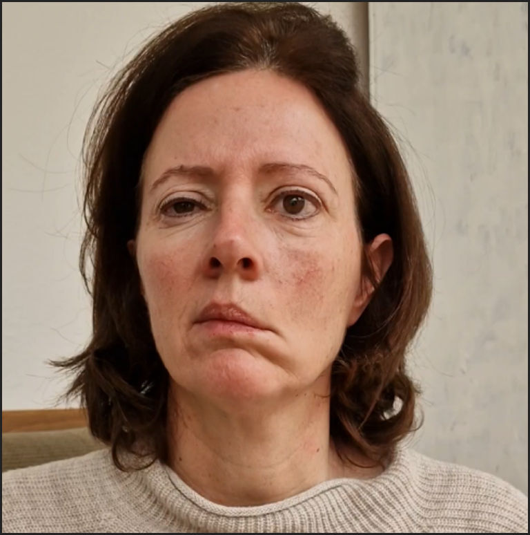
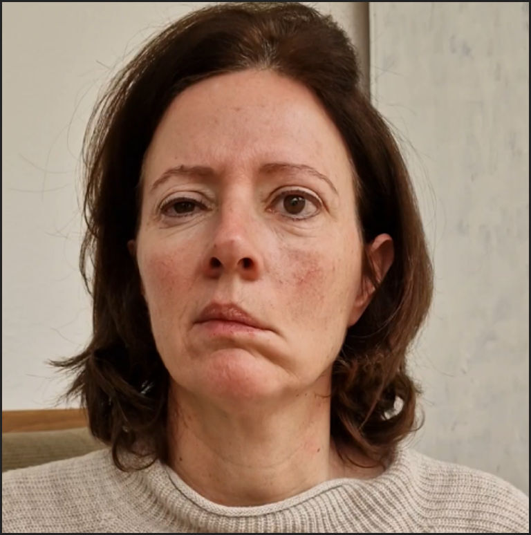
Patient Presentation: 50-year-old female patient was referred by the Clinic of Oral and Maxillofacial Surgery for speech therapy and to improve the function of the lips and cheek muscles due to incomplete facial nerve palsy on the left side following left parotidectomy that was performed 11 months before the patient was referred for myofunctional therapy. The patient presented with visible signs of post-operative peripheral incomplete facial nerve palsy on the left side following parotidectomy. See Figure 1.
The patient underwent intensive therapies including manual stimulation, Novafon Therapy and physiotherapy sessions for over six months, with no significant improvements achieved.
Clinical Presentation: severe facial nerve palsy on the left was characterized by drooping corner of the mouth, raised frown lines, absent forehead wrinkling, incomplete eyelid closure on the left (2 mm) with Sicca symptoms classified as unspecified without documented keratoconjunctivitis, and a still recognizable but smudged nasolabial fold on the left side after removal of the left salivary gland. Inflation of the cheeks was only possible to a limited extent. Sensitivity in the facial area was inconspicuous and without restrictions, with no synkinesis. The patient reported increasing distress regarding facial asymmetry and complained of dry eye symptoms on the left.
The Sunnybrook Facial Grading Score was 25 reflecting severe, incomplete facial paralysis and unrestricted food intake was possible.
Clinically, no contractions were recognizable around the left zygomaticus and risorius muscles. Electromyographically, only isolated potentials could be delineated, which indicated a significantly reduced activation pattern and individual signs of acute denervation. This also applied to the left frontalis muscle.
The findings indicate postoperative effect on the facial nerve, with electromyographically detectable minimal voluntary activity and moderate to marked atrophy of the facial muscles. Needle EMG demonstrated mild to moderate acute and more pronounced chronic denervation signs in the orbicularis oris on the left, with a markedly reduced activation pattern and minimal visible contraction. In the zygomaticus and risorius muscles, no clinical contractions were detectable; EMG showed only isolated potentials with a significantly reduced activation pattern and signs of acute denervation. In the orbicularis oculi and frontalis muscles, only isolated acute denervation signs and minimal voluntary activation were present. Overall, findings indicated a post-operative facial nerve injury with minimal voluntary activity and moderate to severe atrophy of the facial musculature.
Given the residual function of the left facial nerve, augmentation using a masseteric nerve transfer to a zygomatic branch was recommended to preserve and enhance voluntary activation, as spontaneous or natural regeneration was considered unlikely
Treatment Plan Prior to CODONIS vestibular muscle stimulation therapy
Since the patient had severe incomplete facial nerve palsy with no significant improvement seen during a one-year waiting period and because of the residual function of the left facial nerve, the caregivers decided to maintain voluntary activity on the left side and consider a masseteric nerve transfer to a zygomatic branch.
During the first phase of therapy, the focus was on manual activation of the affected facial muscles through gentle stimulation and massage. This included:
-
head and facial stimulation,
-
shoulder loosening massages,
-
chewing exercises,
-
exercises with the Padovan chewing tube,
-
lip-cheek-tongue exercises,
-
cheek inflation to further stimulate the orofacial area of the affected half of the face.
Following the initial phase, a supportive therapy with the Novafon (local vibration therapy) was introduced at home to activate the body’s own mechanisms via targeted tissue stimulation. The therapy was carried out with regular, short-term daily applications. Simultaneously, speech therapy interventions and physiotherapy sessions were held to treat the orbicularis oculi muscle using the gentle myofascial “Anak technique” according to Kurt Ekman Swedish Myofunctional Association (SMA) based upon personal communication.
With a focus on engaging discussions with the patient, regular sessions were held to process the medical history and analyze any new perceptions triggered by the therapy: such as tingling in the cheeks and improved lip movements. The individual progress was recorded in a photo/video documentation and discussed with the patient. In addition, craniosacral therapy was carried out to relax the whole body.
Despite intensive training and high patient motivation, no improvements were achieved in the function of the Orbicularis Oris muscle, the cheek muscles and articulation during the one-year period following the surgery. The progress was slow and limited, and the treatment strategy had to be reviewed. The patient agreed to 'Targeted Muscle Stimulation" using CODONIS ORB device.
Targeted Vestibular Muscle Stimulation using the CODONIS Myofunctional Device for Incomplete Facial Paralysis
The patient consented to CODONIS ORB Vestibular Targeted Muscle Stimulation Therapy with the CODONIS ORB and a therapy plan was prepared that aimed to restore the functionality associated with the balance of the lips and cheek muscles while simultaneously strengthening the orofacial muscles, stabilizing the lower jaw and activating the inside of the (upper) lip in order to reduce the symptoms of paresis.
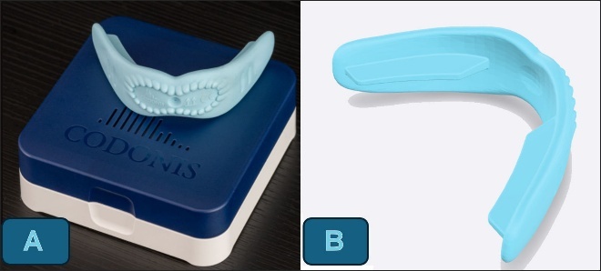
The CODONIS devices, founded by Susanne Codoni, are Swiss-made Class I medical device made with medical-grade TPE-polymer - designed as an intraoral vestibular myofunctional therapy appliance See Figures 2A & 2B. The device rests gently between the lips and teeth to support full mouth closure, stimulates and strengthen the orofacial muscles, encourage correct tongue posture and promote nasal breathing due to complete mouth closure. These devices are recommended for children and adults with speech articulation issues, orofacial and myofunctional disorders.
_codonis_orb_vestibular_muscle_stimulation_device_based_on_kinesiology_principle._the_de.jpeg)
The CODONIS ORB is designed for functional rehabilitation, delivering targeted stimulation to acupressure points within the oral vestibule to activate and align orofacial tissues and postural muscles through a personalized therapy plan. It strengthens orofacial neuromuscular support and promotes airway stability, nasal breathing, facial symmetry, and postural alignment. The device has shown significant improvement in functional rehabilitation in certain medical conditions such as facial paralysis, cerebral palsy, and cleft lip to restore functional integrity and enhance long-term outcomes.
The CODONIS ONE functions as a vestibular myofunctional therapy device that gently activates, coordinates, and harmonizes orofacial muscles. It improves speech articulation, promotes lip seal, nasal breathing, swallowing patterns, tongue positioning and overall oral coordination. Designed for orofacial muscular rehabilitation, it supports clearer speech, balanced facial development and enhanced myofunctional performance across diverse age groups with only a few minutes of use per day.
The intended purpose of the CODONIS ORB in this case was to stimulate, activate, and strengthen the orofacial musculature through targeted vestibular myotherapy. The device is suitable for both professional therapeutic settings and guided home use, with supervision by a trained therapist strongly recommended to ensure correct technique and optimal outcomes.
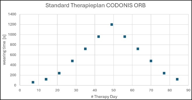
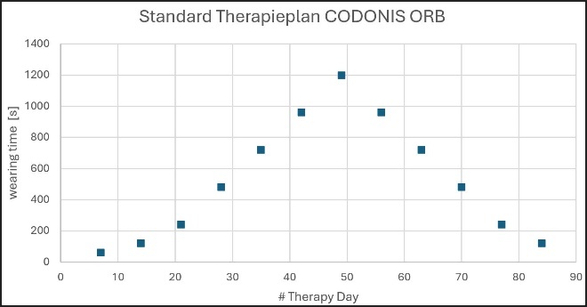
The exercise and therapy plan for this patient was prepared such that the use of CODONIS ORB device continuously increases from 60 seconds from day 0 to day 7, to 120 seconds from day 8 to day 14 gradually increasing up to twenty minutes, then decreases again (in the sense of a bell curve)- rising very slowly and then correspondingly decreasing to provide a structured progressive training therapy with the CODONIS ORB.
In case of incomplete facial paralysis, gradual increase and decrease in stimulation of the orbicularis muscles depending on duration of wear, it helps to reeducate muscle tone without triggering compensatory tension in adjacent muscles. See Figure 3. It supports symmetry restoration and sensorimotor integration, key goals in CODONIS ORB therapy. The slow rise in duration and intensity allows the facial and orofacial muscles to gradually recruit motor units. This mirrors graded activation seen in motor learning — avoiding sudden overload while stimulating proprioceptive feedback loops.
The Gaussian-style progression is not arbitrary — it’s an approach that is biomechanical and neurophysiological designed to optimize activation, prevent fatigue, and sustain long-term improvement in facial motor control.
The standard therapy plan for the CODONIS ORB, was personalized to address the specific needs of the patient.
The therapy was performed as directed by the manufacturer. After washing the hands, the CODONIS ORB was placed in front of the teeth in the oral vestibule and securely positioned with the bite rails of the device. The patient is advised not to clench the teeth but to softly close the mouth. Furthermore, the lips should be closed as well. If this is not possible, the therapist or the patient can assist complete closure of the lips with the fingers to ensure the lips are completely closed with the CODONIS ORB device in. After completing the therapy time, the device is removed from the mouth. The therapy is resumed the next day with the indicated wearing time. The patient was assisted during subsequent therapy sessions online.
Personalized Therapy Plan: Explanation and Figures
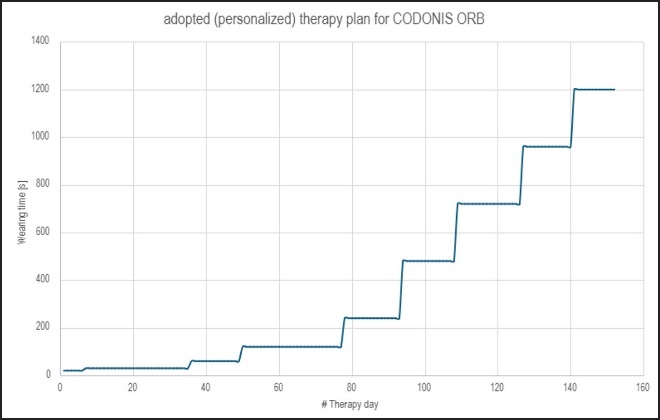
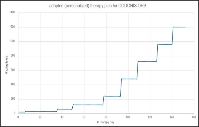
The patient initially presented with significant difficulty in closing the lips, requiring manual assistance during the initial sessions. For this reason, the therapy plan was individualized and based primarily on the patient’s comfort level. The wearing time was introduced for only a few seconds at first and then gradually increased as tolerance improved. This progressive approach was essential to avoid fatigue and ensure compliance. See Figure 4.
Over the course of approximately 160 days, the wearing time was increased stepwise, leading to measurable improvement in lip closure and facial symmetry, as documented in the report. The continuous increase shown in Figure 4 reflects this personalized, adaptive progression. In contrast, Figure 3 represents a standardized reference plan used for typical cases, where wearing time is increased and then decreased in a reciprocal pattern to balance stimulation and rest.
In this case, the standard plan was adjusted to the patient’s specific needs — therefore, the progression in Figure 4 does not mirror Figure 3. The deviation from the standard pattern was intentional and clinically justified to accommodate the patient’s initial limitations and gradual improvement.
Effective results with CODONIS ORB
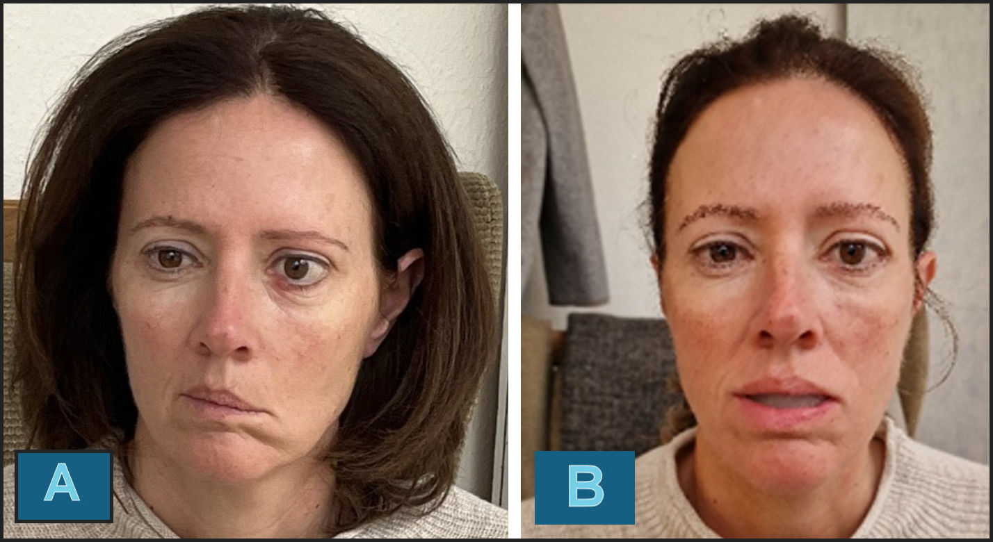
With the use of the CODONIS ORB, significant improvements were observed, and rapid progress was made within three months with gradual increment in wearing time. With visible improvement in the movement and coordination of the lip and cheek muscles, remarkable difference in the shape and function of the lips, intelligibility when distinct improvement in speech with fewer unwanted movements of the lips and cheeks. The patient also experienced a subjective and significant improvement in her aesthetic appearance. See Figures 5A & B.
_initial_presentation_prior_codonis_orb_therapy_b)_initial_therapy_day__patient_wear.jpg)
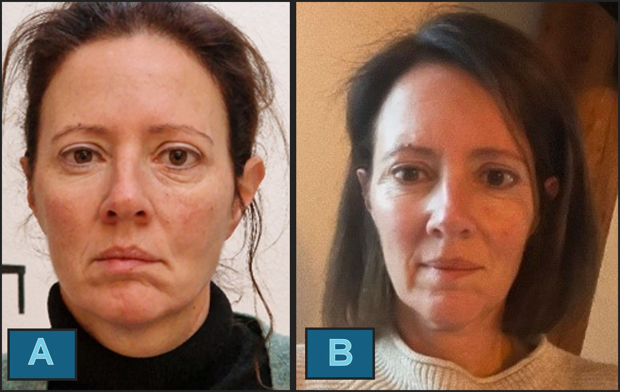
The Sunnybrook Facial Grading Score improved from 25 at baseline to 85 after five months of CODONIS ORB therapy, indicating near-complete restoration of facial symmetry and voluntary movement.
After three months of daily therapy with the CODONIS ORB, almost complete normalization of the facial muscles (facial expressions) was achieved, which also enabled a significant improvement in articulation and speech. See Figure 6A.
_status_after_3_months_of_therapy_with_codonis_orb._b)_status_after_5_months_of_therapy_.jpg)
Further improvements were observed after five months of wearing the CODONIS ORB targeted stimulation device supplemented with intensified training (household work additional massage), from an initial 30seconds to a duration of 1200seconds, a near normal resting position of the facial muscles was evident and improvements in speech were observed. See Figure 6B.
DISCUSSION
The face plays a crucial role in self-perception and social interaction. Facial nerve palsy that effects facial appearance and speech is a potential and known complication of parotidectomy for benign salivary gland tumors, necessitating a comprehensive understanding of its incidence and associated risk factors for effective patient counselling, preoperative planning and post-operative therapies for improved treatment outcome.
Postoperative facial nerve paralysis often results from neurapraxia or axonotmesis following surgical trauma, traction, or edema — particularly in parotidectomy or acoustic neuroma removal. Recovery depends on the degree of nerve injury and neuromuscular responsiveness, with spontaneous improvement typically within six months.17
Postoperative facial nerve dysfunction, primarily involving the seventh cranial nerve, presents a multifactorial challenge affecting both motor and emotional domains. Myofunctional therapy and proprioceptive neuromuscular facilitation (PNF) techniques have shown measurable improvement in facial symmetry, movement quality, and reduction of synkinesis when initiated early.18
The use of the CODONIS ORB in this case described above resulted in remarkable progress during the 5 months of therapy that led to restoration of functionality, reintegration into social life and emotional wellbeing. Myofunctional appliances, in this case targeted muscle stimulation of the orbicularis oris muscle using CODONIS ORB has shown significant improvement in re-establishing facial form and symmetry in the case of partial facial nerve palsy.
The CODONIS ONE and CODONIS ORB represents an intra-oral myofunctional device operating from the oral vestibule, ensuring complete lip closure and enabling lip training through targeted muscle stimulation without external interference from protruding holders or similar appliances that keep the lips open. Vestibular myofunctional appliance activates the entire facial muscle complex through targeted orofacial muscle stimulation, including the orbicularis oculi and orbicularis oris, as well as the connecting musculature, thereby addressing both ocular and oral motor function in an integrated manner. Effective use of the ORB with closed lips for up to two minutes also ensures postural alignment. The use of the CODONIS ORB in this case enabled targeted stimulation to strengthen the affected facial muscles that led to improved lip and cheek function. The patient not only experienced an aesthetic improvement but also increased intelligibility when speaking.
The orofacial system functions as a sensorimotor unit whose stability depends on the coordinated interaction of posture, breathing, and tongue function.19 Consistent with this principle, the positive effects in this case became visible after a short time—already by the 7th week of treatment using the CODONIS ORB device—and continued to increase with ongoing daily training over the following months as presented in Figure 7 and figure 8. The present case aligns with the mentioned use of myofunctional therapy for facial nerve paralysis, demonstrating progressive improvement in orbicularis oculi and oris function, validating the role of structured, non-invasive rehabilitation in facial nerve recovery.
Notably, in this case, the CODONIS ORB therapy was initiated later in the rehabilitation timeline—after conventional physiotherapy and facial retraining had yielded limited improvement. Despite the delayed start, the patient demonstrated measurable functional recovery and aesthetic enhancement. This suggests that targeted vestibular neuromuscular stimulation may retain therapeutic efficacy even when introduced beyond the early intervention window, highlighting its potential as a valuable adjunct in chronic or refractory facial paralysis.
While the outcomes observed in this single case are encouraging, evaluation in a larger patient cohort and further research would provide deeper insight into the functional and neuromuscular effects of the CODONIS vestibular muscle stimulation device.
CONCLUSION
The CODONIS ORB provides a meaningful vestibular myofunctional therapy pathway for patients recovering from invasive procedures such as parotidectomy and its common sequelae, including facial paralysis—offering a non-surgical, accessible alternative in cases where complex and costly interventions like masseteric nerve transfer may not be feasible or desired. It emerges as a compelling, non-invasive alternative to surgical intervention that enhances patients’ quality of life, minimizes the risks typically associated with surgical intervention and helps reduce overall healthcare costs. The successful results of this case report suggest that the CODONIS ORB may serve as a valuable adjunct in the rehabilitation of postoperative facial nerve dysfunction—offering a safe, accessible, and cost-effective therapeutic option that supports functional recovery while reducing the need for further invasive surgical procedures.
Patient perspective
The patient reported a gradual improvement in facial symmetry, lip, cheek and tongue functions and control throughout the therapy sessions. She described the structured progression with the CODONIS ORB as gentle, reassuring and motivating, providing a clear sense of progress and stability during recovery. Over time, she also noted visible enhancement in facial appearance, clarity in speech and smoother functional movement, which positively influenced her confidence and daily interactions.
Informed Consent
The patient provided written informed consent for the clinical procedures described and for the publication of this case report, including all relevant clinical details and images. She specifically agreed that the images may be shown without masking the eye region, as visibility of the orbicularis oculi muscle was necessary to document functional improvement. All identifying information has been removed to preserve confidentiality in accordance with institutional and ethical standards.
Conflict of interest
The authors declare no conflicts of interest related to this case report.
Ethics Statement
This case report describes routine clinical care and does not contain any experimental interventions. According to local regulations, formal ethical approval was not required. Written informed consent for publication of clinical details and images was obtained from the patient.
Reference Integrity Statement
All cited sources have been verified for accuracy and consistency. Internal CODONIS data are clearly identified as unpublished material.
ORCHID ID
Assistant Professor Dr. Serdar Gözler, MDS, PhD: 0000-0002-4899-9517
Assistant Professor Dr. Sarah Qadeer, BDS, MSD: 0000-0002-1389-5996