INTRODUCTION
Dental anxiety affects an estimated 5–30% of adults and approximately one-third of children worldwide and remains a major barrier to oral healthcare.1–6 As a manifestation of sympathetic nervous system activation, it produces the classic physiologic features of the fight-or-flight response: elevated heart rate, increased blood pressure, and heightened apprehension.7,8 These responses lower pain thresholds, increase procedural complications, and create substantial emotional and economic burdens for patients and providers alike.3–6
Current management options, including pharmacologic sedation, behavioral techniques, and complementary approaches such as auricular acupuncture, each carry limitations in cost, side-effect profile, accessibility, or consistency of effect.6,9,10 Most general practitioners lack the training or infrastructure to offer sedation or acupuncture, leaving a practical gap in available anxiolysis options.
Sympathetic modulation offers a physiologically grounded alternative. Stellate ganglion blockade (SGB) interrupts second-order (preganglionic) sympathetic neurons in the cervical chain and has demonstrated anxiolytic effects in post-traumatic stress disorder, also known as PTSD.11–14 The anatomic rationale is well established: interruption of second order (pre-ganglionic) sympathetic neurons in the cervical chain disrupts the feedback loops sustaining pathologic sympathetic tone.11–29 The present study asked whether a simpler, more distal approach-targeting third-order (postganglionic) sympathetic fibers at Erb’s point could produce clinically useful anxiolysis with standard dental equipment. See Table 1.
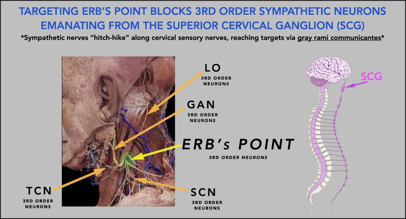
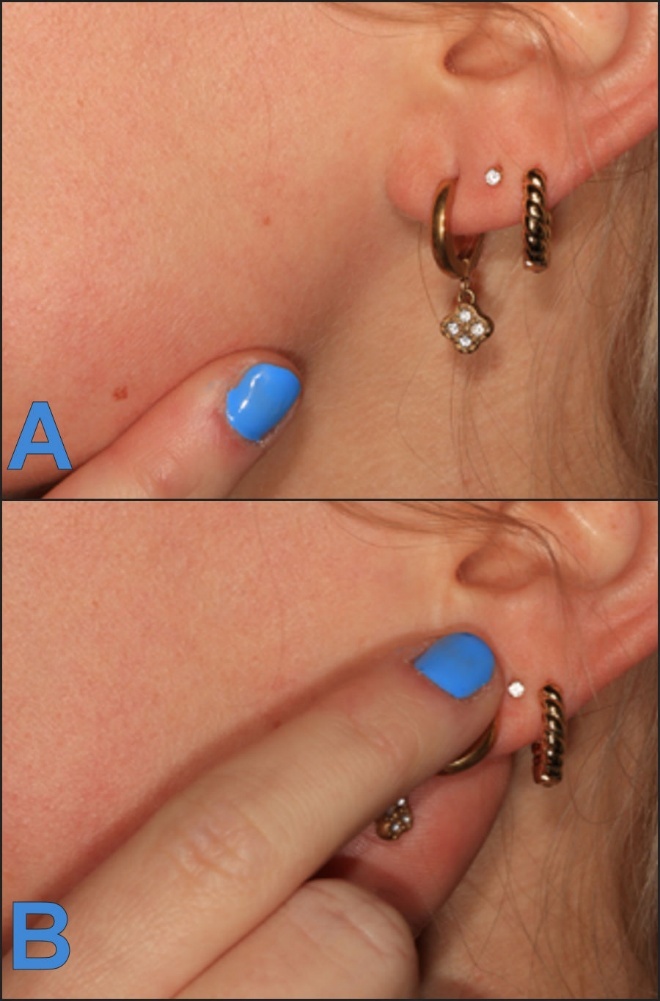
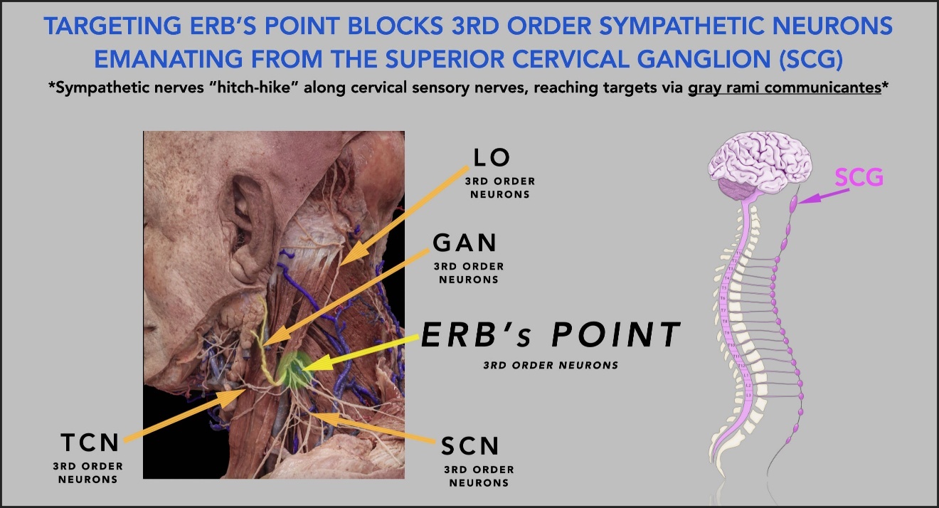
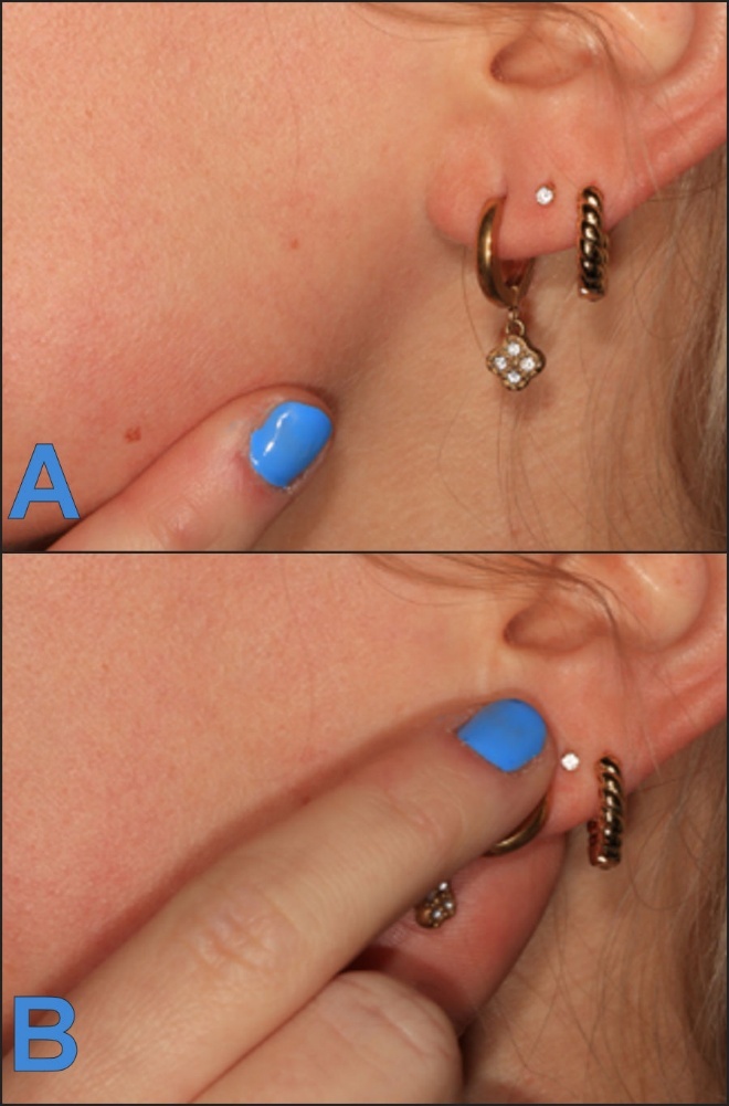
Erb’s point, located at the midpoint of the posterior border of the sternocleidomastoid muscle (SCM), is the emergence site of four cutaneous branches of the superficial cervical plexus; namely the: greater auricular nerve (GAN), transverse cervical nerve (TCN), lesser occipital nerve (LON), and the supraclavicular nerve (SCN). See Figure 1. Postganglionic sympathetic fibers from the superior cervical ganglion travel via gray rami communicantes (GRC) along these cervical sensory nerves.30–32 A superficial injection at this site therefore constitutes a partial cervical plexus block that can interrupt both sensory and sympathetic fibers. Confirmation of anesthesia in the greater auricular nerve distribution (angle of the mandible and ear lobule) served as the clinical endpoint of correct placement of the local anesthetic injected in this study. See Figure 2.
Prior work by the authors has shown that this same injection produces significant temporary reduction of confounding dental cold hypersensitivity in many presenting patients without any intraoral anesthesia, supporting the construct of Sympathetic Dental Hypersensitivity (SDH). The authors posited that neuroinflammation of postganglionic sympathetic C-fibers originating from the superior cervical ganglion (SCG) and traveling with the branches of the superficial cervical plexus (SCP) constitutes a distinct and previously under-recognized endotype of dental pain.33 The dental pulp contains a population of unmyelinated sympathetic fibers arising from the SCG that account for approximately 10% of total pulpal innervation.34 Sympathetically maintained vasoconstriction within the pulpal arteriolar system can produce a compartment-like hypoxic state within the noncompliant dentin shell, driving cold hypersensitivity through a mechanism distinct from the classical hydrodynamic theory.33 That this effect was reproducibly attenuated by local anesthetic deposited at the posterior sternocleidomastoid border, without any intraoral anesthesia, substantially strengthens the case for a sympathetic contribution to intraoral pathology accessible from the cervical level. The present investigation extends that sympathetic anatomic framework to procedural anxiety, postulating that that local anesthetic placed superficially at Erb’s point transiently modulates postganglionic sympathetic fibers traveling with the SCP, providing an accessible, office-based means of reducing dental anxiety through partial interruption of the sympathetic feedback loops that sustain and amplify it.
AIM
To report changes in self-reported anxiety, blood pressure, pulse rate and oxygenation following superficial injection at Erb’s point in anxious dental patients and to provide a foundation for controlled prospective studies.
MATERIALS AND METHODS
This retrospective review examined clinical records from a single private dental practice (NY). Institutional review was deemed unnecessary for a retrospective series of routine procedures. The study was conducted in accordance with the World Medical Association Declaration of Helsinki. Written informed consent for the injection was obtained from all patients or their guardians at the time of treatment.
Inclusion criteria - age >14 years, documented self-reported dental anxiety, and consent for the injection.
Exclusion criteria - allergy to bupivacaine, prior surgery in the distribution of the superficial cervical plexus, patient refusal, or same-day administration of nitrous oxide or benzodiazepines.
Intervention
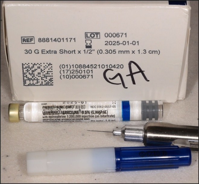
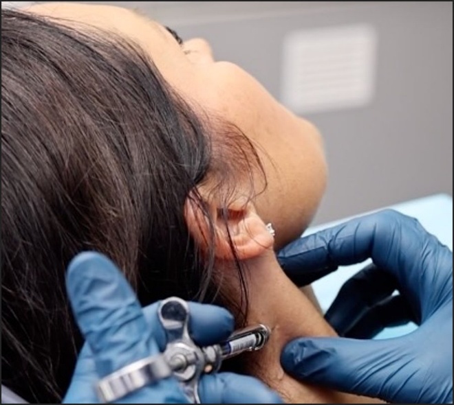
Throughout this study, Marcaine® (bupivacaine HCl 0.5%) with epinephrine 1:200,000 (as bitartrate), 1.8 mL single-dose dental-style cartridge was used as the local anesthetic, whilst an extra short, 30 gauge, ½ inch (13 mm) needle was used to deploy the injectate using a metal aspirating dental syringe. This exact low-profile setup is what enables the minimally invasive, office-based, third-order sympathetic modulation: the tiny volume (~0.5 cc injected from the cartridge) delivered superficially via the fine 30-gauge needle targets the emergence of the superficial cervical plexus branches and hitchhiking gray rami communicantes without deeper spread. See Figure 3.
_with_epinephrine.jpeg)
Pre-injection measurements included VAS anxiety (0–10), blood pressure, pulse, and pulse oximetry. The protocol was as follows:
-
The operative side was confirmed.
-
The skin over Erb’s point on the operative side (right or left since anatomically the superior cervical plexus exists bilaterally) was disinfected with alcohol.
-
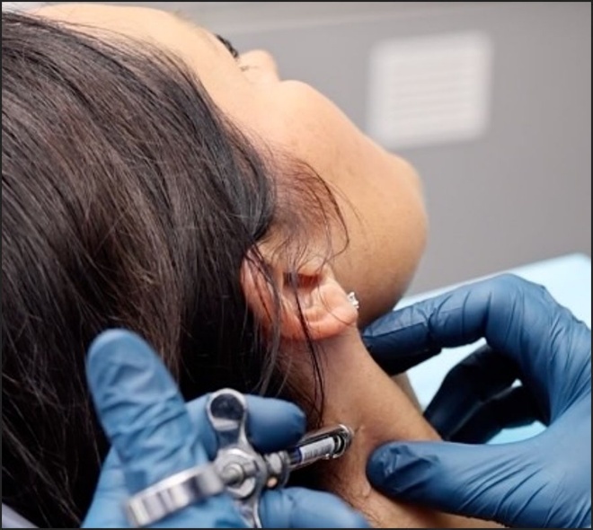
0.4 – 0.8 mL (typically 0.5 mL) of 0.5% bupivacaine with 1:200,000 epinephrine was injected subcutaneously at the midpoint of the posterior border of the sternocleidomastoid using a 30-gauge half-inch needle advanced 3 – 4 mm. See Figure 4.
-
Block success was confirmed by cutaneous anesthesia in the greater auricular nerve distribution on the operative side (angle of mandible-lower lobule of the ear). See Figures 2A & 2B.
-
VAS anxiety, blood pressure, pulse, and pulse oximetry were reassessed 5–10 minutes later, before any intraoral anesthesia.
-
Intraoral anesthesia was administered only after these assessments.
No sham or comparison injections were used. Patients were aware that the injection was intended to assist with anxiety via the consent form. However, their subjective VAS pre-injection anxiety level was documented before they became aware of the potential usage and intention of the Erb’s block.
Statistical Analysis
Data were tested for normality (Shapiro-Wilk and Shapiro-Francia). The Wilcoxon Signed-Rank test was used for paired comparisons. Cohen’s d was calculated for effect size.
RESULTS
Paired data were available for all 132 patients for blood pressure. Pulse rate data were missing for 10 subjects (n = 122) and post-injection anxiety scores for four subjects (n = 128).
The mean systolic blood pressure, diastolic blood pressure, and pulse rate each declined significantly after injection (p < 0.05), although effect sizes were all small. See Table 1. The mean pulse oximetry was unchanged as expected after the short time between pre and post injection. No adverse events were recorded.
Self-reported anxiety decreased significantly. The mean reported VAS anxiety score (0 – 10) fell from 3.88 (±2.56) to 1.57 (±1.58), a 60% reduction (Wilcoxon signed-rank test, p < 0.00001) with a large effect size (Cohen’s d = 1.086). Pain scores did not change significantly because there was very little pain associated with this group of routine, non-TMD dental patients. Therefore, the reports of reduced anxiety could not be attributed to any reduction in pain. See Table 2.
Most patients found the neck injection less objectionable than typical intraoral injections. Many described a generalized, whole-body sense of relaxation 3 - 5 minutes after the unilateral block. The anxiolytic effect appeared to last for the duration of the local anesthetic (approximately 4–6 hours), and several patients later reported surprisingly improved sleep quality on the night of the Erb’s point injection. The next paragraph compares and contrasts potential local anesthetic choices employed by the authors for the Erb’s block, though throughout this study, bupivacaine with 1:200,000 epinephrine was utilized.
DISCUSSION
In this retrospective series, superficial injection at Erb’s point was associated with significant reductions in self-reported anxiety and modest decreases in blood pressure and pulse. The anatomic rationale is coherent: postganglionic sympathetic fibers traveling with the superficial cervical plexus at Erb’s point are accessible to local anesthetic, producing downstream effects on anxiety and hemodynamics that parallel those reported with stellate ganglion block,11–29 albeit at a more distal (third order/post-ganglionic) level.
Alternative explanations cannot be excluded. Placebo and expectation effects on subjective anxiety scales are well documented in the literature. Though unlikely, sensory anesthesia of the superficial cervical plexus itself may contribute to the findings. Although anticipatory anxiety naturally declines once treatment begins, patients had yet to undergo their scheduled dental operative treatment procedures following both pre-block and post-block assessments. Without a sham injection or untreated control, a specific sympathetic mechanism cannot be isolated. With all things considered, the data are consistent with the sympathetic hypothesis, but the findings using the protocol within this retrospective case series do not confirm it.
All pre-injection measurements were obtained before patients were informed of the possibility of the neck injection, and all post-injection measurements were obtained before intraoral anesthesia, preserving the original anxiety-provoking context (be it the “shot”, the noise, the smells, and/or the impending fear of the upcoming dental procedure).
The systemic signal embedded in the vital sign findings warrants consideration beyond the immediate question of procedural anxiety. A reduction in both systolic and diastolic blood pressure following injection of half a milliliter of local anesthetic containing epinephrine at a superficial cervical site implies a degree of systemic vasodilation. The physiologic significance of this observation extends to a broader question: what consequences might chronic sympathetic overdrive carry at the tissue level in the orofacial environment, and has that question been systematically examined?
Our prior work demonstrating temporary resolution of dental cold hypersensitivity with the identical injection supports a clinically relevant role for cervical sympathetic fibers in intraoral pathology. The present anxiolysis findings and those earlier hypersensitivity findings arise from anatomically equivalent injections and together constitute convergent preliminary evidence.
Sympathetic Tone and the Orofacial Environment
There is no anatomic or physiologic basis for the facial and orofacial tissues to be exempt from the consequences of sustained sympathetic activity that are well established elsewhere in the body. Chronic sympathetic overdrive produces vasoconstriction, impairs local tissue perfusion, delays wound healing, reduces effective delivery of pharmacologic agents to target tissues, and is implicated in chronic pain states including Complex Regional Pain Syndrome, Type 1.35,36 Presentations in orofacial patients attributed to structural, infectious, or idiopathic causes may in some proportion reflect sympathetic dystonia that has not been included in the differential diagnosis.
The clinical relationship between psychological stress and orofacial pathology is familiar. Patients under chronic psychological stress present with masticatory muscle hypertonicity, bruxism, temporomandibular dysfunction, and mucosal changes, all of which are mediated at least in part through sympathetic nervous system activation. The extent to which sustained sympathetic activity generates end-organ consequences in the orofacial environment, beyond the recognized acute stress response, has not been subjected to systematic investigation.
The work of Yiannios and colleagues provides the most direct available evidence that sympathetic innervation via the superficial cervical plexus participates in clinically significant dental pathology.33 In a retrospective series of 194 patients with confounding dental cold hypersensitivity, injection of local anesthetic at the posterior border of the sternocleidomastoid, targeting the greater auricular nerve at the point of its emergence from the deep cervical fascia, produced significant temporary reduction of cold sensitivity in 129 subjects (66.5%) without any intraoral trigeminal anesthesia, and produced complete or near complete resolution in 58 subjects (29.9%).33 As noted above, this injection site is anatomically the superficial portion of Erb’s point, and the technique is identical to that employed in the present series. The authors proposed that neuroinflammation of postganglionic sympathetic C-fibers originating from the superior cervical ganglion, traveling with the branches of the superficial cervical plexus, constitutes a discrete endotype of dental cold hypersensitivity they designated Sympathetic Dental Hypersensitivity.33 The proposed mechanism involves sympathetically maintained vasoconstriction within the pulpal arteriolar system, producing a compartment-like hypoxic state within the noncompliant dentin shell, with cold hypersensitivity as a downstream consequence.33 That this effect was reproducibly attenuated by subcutaneous injection at the posterior sternocleidomastoid border, without any intraoral trigeminal anesthesia, substantially strengthens the case for a sympathetic component to intraoral pathology that is accessible from the cervical level.
The two observations, procedural anxiolysis in the present series and attenuation of dental cold hypersensitivity in the series of Yiannios et al,33 arise from injections that are anatomically equivalent in site and technique and differ only in the clinical question examined. Taken together, they constitute convergent preliminary evidence for a clinically meaningful role of the superficial cervical sympathetic distribution in orofacial pathology.
Candidate Conditions for Future Investigation
Several orofacial and dental conditions represent plausible candidates for sympathetically mediated or sympathetically amplified pathology. Each constitutes a discrete line of future inquiry.
-
Dental Cold Hypersensitivity and the SDH Endotype. A substantial proportion of patients with confounding dental cold hypersensitivity appear to have a sympathetic etiologic component accessible at the level of the superficial cervical plexus. A cervical plexus injection at Erb’s point may offer a complementary diagnostic and therapeutic approach in patients with the SDH endotype (33).
-
Chronic Orofacial Pain and Complex Regional Pain Syndrome. Complex Regional Pain Syndrome Type 1 is a recognized consequence of sympathetic dysregulation in the extremities. Whether an analogous process occurs within the trigeminal distribution—contributing to atypical facial pain, burning mouth syndrome, or refractory odontogenic pain without identifiable structural cause—has not been adequately studied. A repeatable superficial cervical plexus block at Erb’s point offers a practical clinical probe for this hypothesis.
-
Pulpitis and Unexplained Tooth Sensitivity. Approximately 10% of pulpal nerve fibers are unmyelinated postganglionic sympathetic C-fibers arising from the superior cervical ganglion. These fibers regulate pulpal blood flow; under conditions of chronic sympathetic activation, they can drive vasoconstriction and a hypoxic cascade within the pulpal compartment.34 Whether the Erb’s blockade produces measurable attenuation of symptomatic pulpitis or unexplained heat sensitivity is directly testable.
-
Alveolar Osteitis. The pathophysiology of alveolar osteitis remains incompletely resolved. Sympathetically mediated vasoconstriction impairing alveolar perfusion and clot stability is a hypothesis that has not been systematically evaluated. Anxious patients presenting with elevated sympathetic tone at the time of extraction may be at higher risk through a perfusion-mediated mechanism.
-
Wound Healing and Post-Surgical Recovery. Sympathetic vasoconstriction restricts delivery of oxygen, inflammatory mediators, and pharmacologic agents to healing surgical sites. Regional sympathetic modulation with the technique described here, applied before or after oral surgery, could potentially improve the local healing environment without systemic pharmacologic intervention.35
-
Chronic Infection and Periodontal Disease. Tissue perfusion is a determinant of local immune competence. Chronic sympathetically mediated vasoconstriction in the periodontal tissues could impair the local immune environment and contribute to infection persistence and periodontal breakdown. An epidemiologic association between chronic psychological stress and periodontal disease is established;37,38 the tissue-level mechanism warrants examination under a sympathetic framework.
-
Unexplained Masticatory Muscle Dysfunction. Masticatory muscle spasm, hypertonicity, and chronic myofascial pain in the absence of identifiable temporomandibular joint structural pathology are common and clinically challenging. Sympathetic-sensory coupling in craniofacial nociception has been proposed as a contributing mechanism.39,40 Empiric use of the Erb’s blockade in such cases represents a low-risk clinical trial.
-
Explaining the Stress–Orofacial Pain Connection. The superior cervical ganglion regulates blood flow not only to the dental pulp but also to the muscles of mastication, temporomandibular joints, and other cephalic structures. Sympathetically mediated mechanisms may help explain the frequently observed but poorly understood link between psychological stress and orofacial pain.
Proposed Next Steps
This series is a preliminary clinical observation. The findings justify a structured program of investigations beginning with the anxiolysis question and extending toward the broader sympathetic hypotheses outlined above.
The immediate research priority is a randomized, sham-controlled trial of superficial cervical plexus injection at Erb’s point for dental anxiety, incorporating a blinded outcome assessor, a comparison injection at an alternative cervical site, pre-specified primary endpoints including both VAS anxiety scores and objective vital sign parameters (e.g., salivary cortisol). Such a design could distinguish a site-specific effect from nonspecific responses to cervical injection.
Concurrently, pilot series applying the same technique and outcome measurements to patients with unexplained masticatory muscle dysfunction, the SDH endotype as defined by Yiannios and colleagues,33 and impaired post-surgical healing could generate preliminary data to support adequate confirmatory trials.
Longer-term investigative questions include whether baseline sympathetic tone, quantifiable through heart rate variability, skin conductance, or thermographic imaging of facial vasomotor patterns, predict a therapeutic response to superficial cervical plexus blockade. Additional questions include whether pre-procedural injection at Erb’s point alters post-operative infection rates, healing timelines, or analgesic requirements following oral surgery, and whether a subgroup of patients with chronic orofacial pain exists in whom sympathetic dysregulation is a primary rather than contributing pathophysiologic mechanism.
LIMITATIONS
This was a retrospective, uncontrolled series from a single operator (NY). No sham or comparison injection was used. Anxiety was measured only by subjective VAS without blinded assessment or objective autonomic measures. The proposed sympathetic mechanism, while anatomically coherent, remains unconfirmed by direct evidence in this study.
CONCLUSION
This pilot series describes a simple, low-risk, office-based injection at Erb’s point associated with significant reductions in dental anxiety and modest improvements in vital signs. The technique requires no special equipment, takes less than one minute to accomplish, and is readily performed in a standard dental setting. The authors propose the term Erb’s Sympathetic Anxiolytic Technique (ERB’sSAT) to describe the Erb’s block for anxiolytic applications. Table 3 compares and contrasts the preganglionic SGB blockade to the postganglionic ERB’sSAT blockade.
Although the effect size for anxiety reduction was large, the absence of a control condition prevents definitive mechanistic conclusions. The anatomic rationale involving the sympathetics and the convergent findings with prior cold-hypersensitivity work support further investigation. A randomized, sham-controlled trial is the necessary next step. The clinical opportunity exists in every dental practice; the barrier to rigorous study is low.
ABBREVIATIONS
SNS, sympathetic nervous system; SCG, superior cervical ganglion; SCP, superficial cervical plexus; SGB, stellate ganglion block; GRC, gray rami communicantes; VAS, verbal annunciated scale; GAN, greater auricular nerve; ERB’sSAT, Erb’s Sympathetic Anxiolytic Technique; SDH, Sympathetic Dental Hypersensitivity.
FUNDING
No funding was sought or received for this research study.
ACKNOWLEDGMENTS
We would like to thank Mrs. Kennedy McDowell, RDA, Mr. Carson Cassetty, BA, RDA, and Ms. Sydney Pawlowski BS, RDA for clinical data gathering and organizational assistance.
DISCLOSURES
John Radke is Chairman of the Board of BioResearch Associates, Inc., and receives no sales-related incentive. The MP and NY declare no conflicts.
CORRESPONDENCE
Mark A. Piper DMD, MD
Email: drpiper@pipereducation.org
Nick C. Yiannios DDS
Email: drnick@drnickdds.com
DECLARATION OF GENERATIVE AI USE
The AI tool Claude (Anthropic) was used in the preparation of this manuscript for the following purposes: iterative drafting and structural reorganization of manuscript sections, editing for scholarly register and consistency of voice, identification and elimination of informal or colloquial phrasing, and formatting of the reference list in Vancouver style. All scientific content, clinical observations, data, mechanistic hypotheses, and conclusions originate with the authors. No AI tool was used for data analysis, data interpretation, or generation of study findings. All AI-assisted text was reviewed, edited, and approved by the authors, who take full responsibility for the accuracy and integrity of the published work.