Introduction
Temporomandibular disorders (TMDs) are multifactorial conditions involving the temporomandibular joints, masticatory muscles, and associated structures. Myalgia and myofascial pain are common pain-related TMD presentations. The Diagnostic Criteria for Temporomandibular Disorders (DC/TMD), formulated at a consensus conference from the invalid RDC/TMD, provides an unvalidated clinical framework for diagnosing common TMD conditions. For pain-related TMD, diagnosis is based on symptom history and standardized examination, including pain modified by jaw movement, function, parafunction and the reproduction of familiar pain during palpation or mandibular movement. The DC/TMD framework does not use imaging, surface electromyography (sEMG), digital occlusal analysis (T-Scan), joint vibration analysis (JVA), mandibular tracking, or bite-force measurement as standalone diagnostic criteria for evaluating myofascial pain patients.1,2
Subsequent DC/TMD translation and implementation literature continues to support DC/TMD as a contemporary clinical framework for standardized assessment of common temporomandibular disorders.3 This supports the diagnostic framing used in the present case: the myofascial pain diagnosis was clinical, while the biometric recordings were used to characterize function under defined loading conditions.
The relationship between dental occlusion and TMD remains controversial. Contemporary literature does not support a simplistic model in which occlusion alone is considered a universal or primary cause of TMD. Manfredini et al. concluded that available association studies do not support a clinically relevant disease-specific association between dental occlusion and TMD.4 Similarly, Al-Ani summarized the long-standing controversy and cautioned against reducing TMD to an occlusal disease model.5 Unfortunately, these conclusions were reached without objective functional measurements and in the context of a longstanding failure to incorporate more than three decades of published occlusal biometric literature. This omission is consequential because it has shaped the interpretation of occlusion in TMD by allowing conclusions to be drawn without adequate consideration of objective data on occlusal loading, timing, mandibular function, and neuromuscular recruitment. Experimental occlusal-interference studies have shown that altered occlusal input may influence motor behavior without reliably producing TMD symptoms in all subjects.6,7 Therefore, the clinically relevant question may not be whether occlusion universally causes TMD, but whether alteration of the occlusal loading condition systematically modifies neuromuscular recruitment and force expression in a given patient.
Recent cross-sectional evidence also supports caution when interpreting static occlusal classification. In generally healthy young adults with Class I and Class II malocclusion examined using DC/TMD forms, Gałczyńska-Rusin et al. found no significant association between malocclusion class and masticatory muscle pain.8 These findings reinforce the distinction between static malocclusion categories and patient-specific functional loading behavior.
Surface electromyography has been studied for many decades as a method for evaluating masticatory muscle activity in pain-related TMD. A recent systematic review reported that patients with pain-related TMD may demonstrate altered masticatory muscle activity, including task-dependent differences during rest and clenching, while also emphasizing heterogeneity and limited standalone diagnostic utility.9 Consequently, sEMG is best interpreted as an adjunctive functional assessment rather than a replacement for established clinical diagnostic criteria.
Additional recent evidence further supports the task-dependent nature of masticatory muscle recordings. Lin et al. evaluated masseteric activity during maximum mouth opening in healthy volunteers and TMD patients with limited mouth opening, myofascial pain, and/or disc displacement; their findings showed subgroup- and task-specific differences in muscle activity rather than a single diagnostic EMG pattern, further supporting the interpretation of sEMG as a functional descriptor rather than a standalone diagnostic criterion.10
Digital occlusal analysis and synchronized electromyography have also been used to evaluate relationships among occlusal timing, force distribution, and masticatory muscle activity. The Disclusion Time Reduction (DTR)/immediate complete anterior guidance development (ICAGD) literature has reported that T-Scan and EMG-guided occlusal therapy may influence symptoms and muscle function in selected occluso-muscular TMD populations.11–18 Although these technologies do not replace established clinical diagnostic criteria, they support the concept that occlusal force/timing relationships and muscle activity may be evaluated simultaneously under defined functional conditions.
Innobyte adds a separate bite-force measurement dimension. Ustrell-Barral et al. reported that the Innobyte system provides reliable maximum bite-force measurements in healthy adults with natural dentitions and provided reference values for maximum bite force.19 In the present example, Innobyte was not used as a diagnostic test for myofascial pain, but rather as a controlled interocclusal loading challenge to determine whether modification of the occlusal environment altered masticatory muscle recruitment.
The present case example describes a patient with clinically diagnosed myofascial pain in whom sEMG, T-Scan, and Innobyte recordings demonstrated a marked occlusion-dependent alteration in masticatory muscle recruitment and force expression.
An Example
An adult male patient presented for evaluation of head, neck, jaw, and facial pain symptoms. The chief complaints included jaw pain, chewing pain, jaw clicking, jaw locking-type symptoms, neck pain, morning head pain, ear symptoms, dizziness, and frequent heavy snoring. The patient reported that chewing and hard foods aggravated symptoms, while analgesic medication provided partial relief.
Relevant medical history included hypertension, chronic pain, sleep apnea/CPAP history, neck and shoulder pain, and use of medication for pain management. Airway screening identified increased neck circumference, high tongue posture, Mallampati Class IV appearance, enlarged tongue, absent tonsils, and history suggestive of sleep-disordered breathing. These findings were considered clinically relevant comorbidities and potential modifiers of pain perception, fatigue, and neuromuscular function, potentially contributing to symptom amplification and reduced physiologic recovery; however, they were not used as primary diagnostic criteria for myofascial pain.
Clinical examination demonstrated widespread tenderness of the masticatory and associated cervical musculature. Tenderness was recorded bilaterally in the temporalis, masseter, digastric, medial and lateral pterygoid regions, sternocleidomastoid, trapezius/shoulder region, temporal tendon, lateral TMJ capsule, and posterior joint space. The patient’s symptom history, function-related pain, and reproduction of pain during palpation supported a diagnosis of masticatory and cervical myalgia/myofascial pain consistent with DC/TMD principles.
Mandibular range of motion was functionally preserved. Clinical notes recorded maximum opening of approximately 65 mm, right lateral movement approximately 6 mm, left lateral movement approximately 6 mm, and protrusion approximately 9 mm. Jaw Tracker recordings demonstrated vertical opening of approximately 45-46 mm in the captured functional recordings, with preserved opening capacity and mild functional asymmetry. The difference between clinical maximum opening and Jaw Tracker-recorded opening was interpreted as task- and recording-condition specific rather than evidence of a fixed limitation of mandibular range. (Table 1.)
Materials and Methods
Example case design and diagnostic standard
This manuscript is a retrospective, single-patient clinical example case based on de-identified records obtained during routine clinical assessment. No experimental intervention was performed for research purposes. The report was prepared to describe the relationship between clinical findings and adjunctive functional recordings obtained under defined occlusal loading conditions.
The diagnosis of masticatory and cervical myalgia/myofascial pain was made clinically using history and examination findings consistent with DC/TMD principles. The diagnostic standard was symptom history, pain modified by jaw function or parafunction, and reproduction of familiar pain during examination of the masticatory and associated cervical musculature. Instrumental recordings were not used as standalone diagnostic criteria for myofascial pain.
Clinical examination and conventional occlusal record
Clinical examination included review of chief complaints, aggravating and relieving factors, relevant medical and sleep-airway history, mandibular range of motion, temporomandibular joint screening, and bilateral palpation of the masticatory and associated cervical musculature. Recorded muscles and regions included the temporalis, masseter, digastric region, medial and lateral pterygoid regions, sternocleidomastoid, trapezius/shoulder region, temporal tendon, lateral TMJ capsule, and posterior joint space.
Clinical intraoral photographs and 100 µm articulating-paper markings were obtained as a conventional visual record of tooth contact location. Articulating paper was not used to quantify occlusal force, occlusal timing, right/left force balance, or muscle activity. Force magnitude and timing were interpreted only from digital occlusal analysis, and bite force in Newtons was interpreted only from Innobyte recordings.
Instrumentation and software configuration
The functional assessment used commercially available clinical recording systems. BioPAK software version 9.1.0 Beta 17 (BioResearch Associates, Inc., Milwaukee, WI, USA) was used for BioResearch modules, including JVA, EMG-8, JT-3D, bite-registration, mastication EMG, and T-Scan Link integration. Digital occlusal analysis was performed using T-Scan 10 software version 10.32 (Tekscan, Inc., Norwood, MA, USA) with a T-Scan Novus USB handpiece (model DH-1) and T-Scan Novus Large sensor. Maximum bite-force measurement was performed using the Innobyte system (Kube Innovation Inc., Montreal, QC, Canada). The specific software and device descriptions are provided to allow readers to identify the measurement systems used and to distinguish the different variables generated by each device.
Software versions and device identity were documented from the clinical systems available at the time of review. All systems were operated according to the manufacturers’ clinical software and hardware configuration settings available in the installed systems. No research-specific calibration, signal filtering, normalization, engineering validation, or independent recalibration was performed beyond routine manufacturer-guided clinical use. The report therefore describes the clinical measurements displayed and captured by the manufacturers’ systems during routine assessment, rather than independently validated engineering measurements.
Surface electromyography
Surface electromyography (sEMG) was recorded with BioEMG III/BioPAK using the EMG-8 module under the manufacturer’s clinical software/hardware settings. sEMG records electrical activity from the selected muscles through surface electrodes and reports muscle activity in microvolts (µV). For this example case, the clinically relevant comparison focused on the elevator-muscle channels: right anterior temporalis (TA-R), left anterior temporalis (TA-L), right masseter (MM-R), and left masseter (MM-L). The manuscript reports the displayed clinical output values from the BioPAK/BioEMG system; the values were not independently normalized to a maximum voluntary contraction reference and were not treated as normative diagnostic thresholds. General surface-electrode acquisition principles, including attention to electrode placement and skin preparation, are consistent with established sEMG sensor and sensor-placement recommendations.20
The primary sEMG outcome was mean elevator-muscle activity, calculated as the arithmetic mean of TA-R, TA-L, MM-R, and MM-L during the selected clench recording. The reported values therefore represent recorded surface electrical activity during the task; they should not be interpreted as direct force values, pain scores, or diagnostic thresholds for myofascial pain.
Two sEMG clench conditions were compared: clenching into the natural dentition and clenching with the Innobyte interocclusal device positioned between the teeth. The purpose of this comparison was to determine whether changing the interocclusal loading environment altered elevator-muscle recruitment. The same muscle groups and the same reporting units were used in both conditions.
Digital occlusal analysis
Digital occlusal analysis was performed using T-Scan 10. The T-Scan system uses a thin intraoral pressure-sensitive sensor connected to software that records the sequence, timing, and relative distribution of occlusal contacts during mandibular closure and clenching. In this case example, T-Scan was used to document the natural intercuspal loading pattern during clench into the dentition.
For this recording, the T-Scan Novus USB handpiece (model DH-1) was used with a Large T-Scan Novus sensor (Large 2002 sensor; Tekscan, Inc., Norwood, MA, USA). The manufacturer describes the Novus handpiece as the hardware interface that captures data from the dental sensor and transfers it to the T-Scan software; the sensor support clips onto the handpiece, and the thin pressure-sensitive dental sensor is inserted into the support. The Large sensor was selected to fit the patient’s arch size and to cover the dental arch during closure. Manufacturer labeling identifies T-Scan Novus sensors as available in small and large sizes intended for intraoral bite-force readings, wafer-thin, high-resolution Sensel-based sensors. The package used in this example case was a large sensor package, marked single-patient use.21,22
The principal T-Scan variables reported were right-side relative force percentage, left-side relative force percentage, dominant loading region, and the visual force-time pattern during the selected recording. The T-Scan Novus handpiece records dental sensor data at a standard rate of 175 Hz, with higher acquisition available in Turbo Mode according to manufacturer product information.21 T-Scan percentages are relative values within the recording and represent the distribution of sensor-recorded occlusal load across the arch. They are not absolute bite-force values in Newtons and should not be compared numerically with Innobyte Newton measurements.
The T-Scan handpiece, sensor, and software were used according to the manufacturer’s clinical settings and normal operating workflow. Sensor conditioning, sensitivity selection, and recording parameters were governed by the installed T-Scan 10 software/hardware configuration. The percentages reported in this manuscript are therefore the software-displayed relative force distribution values from the selected clinical recording, not independently calibrated absolute-force measurements.
The T-Scan recording was interpreted as a functional occlusal loading record. It was used to identify whether natural intercuspation produced a balanced or asymmetric loading platform and whether load was concentrated anteriorly or posteriorly. Sensor data were interpreted clinically together with intraoral findings and sEMG/Innobyte data rather than as an isolated diagnostic test.
Innobyte bite-force measurement and interocclusal challenge
Maximum bite-force testing was performed with the Innobyte system (Kube Innovation Inc., Montreal, QC, Canada), using the bilateral mouthpiece configuration connected to the handheld Innobyte display unit. Innobyte is a dental bite-force measurement device intended for quantitative evaluation of maximal bite strength. It reports force in Newtons (N), not as a relative percentage. In this example case, the device was used both to quantify force-generating capacity and to provide a standardized interocclusal loading challenge.
The intraoral measuring element was the Innobyte bilateral mouthpiece (INNOMP03). According to the manufacturer’s user guide and technical specifications, the mouthpiece is the applied part of the device and is placed between the maxillary and mandibular teeth during biting.23 A bilateral mouthpiece configuration was selected to provide a standardized loading platform during force testing. Manufacturer-reported overall mouthpiece dimensions are approximately 101 x 63 x 32 mm (length x width x height), with an approximate weight of 56 g. Because the device introduces a defined interocclusal platform, recordings were obtained under standardized loading conditions rather than direct tooth-to-tooth intercuspation.
The manufacturer describes the mouthpiece as containing a fluid medium. When force is applied by the teeth, the force is transmitted through the fluid and read by internal sensors; each mouthpiece is calibrated to correlate pressure with force in Newtons. The manufacturer reports a measurement range of 0 to 2000 N, data acquisition up to 100 Hz, and accuracy within 5% of full scale under the specified operating conditions.23 The bilateral mouthpiece displays the maximum total bite-force value on the superior row of the display and the maximum left- and right-side bite-force values on the inferior row, relative to the orientation of the mouthpiece.
For recording, the mouthpiece was covered with a new disposable cover, inserted using the handle, and positioned according to the manufacturer’s instructions: the maxillary central incisors were placed against the protruding anterior stop and the bilateral cheek guards were positioned against the molar regions. The patient was instructed to bite maximally on the mouthpiece for approximately 1 second. Consecutive recordings were reset using the device interface before the next bite. The manufacturer recommends three measurements with up to 10 seconds of rest between measurements23; in the present example, the reported values were summarized descriptively from the available static bite and repeated-clench recordings.
The Innobyte device was used according to the manufacturer’s normal operating instructions and factory-calibrated mouthpiece/display workflow. No research-specific modification of the mouthpiece, bite insert, software, or hardware settings was performed. Recorded values represent the force values displayed by the Innobyte system in Newtons under routine clinical acquisition conditions.
Innobyte variables reported in this manuscript were total bite force (N), right-side bite force (N), and left-side bite force (N). Static bite and repeated-clench recordings were summarized. These measurements represent absolute force values and should not be interpreted as equivalent to the relative force-distribution percentages reported by T-Scan.
The Innobyte-supported clench was interpreted as an interocclusal challenge because the 101 x 63 x 32 mm bilateral silicone mouthpiece necessarily altered several variables simultaneously: vertical dimension, tooth contact input, periodontal afferent input, mandibular stabilization, contact distribution, and patient load tolerance. The Innobyte recordings were interpreted as adjunctive functional measurements and were considered together with clinical examination findings, digital occlusal analysis, and sEMG recordings rather than as an isolated diagnostic test.
Mandibular movement analysis
Mandibular movement was recorded with the JT-3D Jaw Tracker system within BioPAK. Jaw tracking records mandibular movement during opening, closing, and excursions. Reported variables included vertical opening, anterior/posterior movement, lateral deviation or deflection, and maximum slant or path-related movement values, expressed in millimeters. In this case example, Jaw Tracker data were used to document functional mandibular movement capacity and to determine whether the presentation was suggestive of a fixed closed-lock pattern.
Clinical maximum opening and Jaw Tracker-recorded opening were interpreted separately. Clinical range of motion represented chairside maximum opening, whereas Jaw Tracker values represented the movement captured during the selected recorded task. Differences between these values were interpreted as task- and recording-dependent rather than as contradictory measurements.
Joint vibration analysis
Joint vibration analysis was performed using BioJVA within BioPAK. JVA records vibrations associated with mandibular opening and closing and displays vibration amplitude and frequency characteristics from the temporomandibular joints. The primary variable referenced in this report was the selected-cycle total integral, with attention to whether a prominent vibration signature was present.
JVA was used as contextual functional information only. A low or absent vibration recording in a selected cycle does not exclude all intra-articular pathology, disc displacement, degenerative change, or intermittent joint sound. JVA findings were therefore interpreted cautiously and were not used to establish or exclude the clinical diagnosis of myofascial pain.
Recording conditions and task comparison
All occlusal and bite-force measurements were made with the patient seated upright in a standardized supported posture. The patient was positioned facing forward, with the back and neck supported and the occlusal plane approximately parallel to the floor. The legs and arms were kept uncrossed. This posture was used to reduce avoidable postural, cervical, shoulder-girdle, and fascial strain during occlusal loading records and to improve consistency across the natural-clench, T-Scan, sEMG, and Innobyte recording conditions.
All instrumental recordings were obtained during the same clinical assessment as part of routine functional documentation. The key comparison was between two loading conditions: (1) natural clench into the patient’s dentition and (2) clench with the Innobyte interposed between the teeth. The natural-clench condition was used for the T-Scan force-distribution recording and for baseline sEMG. The Innobyte-supported condition was used to record bite force in Newtons and to determine whether elevator-muscle recruitment changed when the interocclusal loading condition was modified.
The variables compared across conditions were not treated as interchangeable. sEMG measured electrical muscle recruitment in µV; T-Scan measured relative intra-arch force distribution and timing; Innobyte measured absolute bite force in Newtons; Jaw Tracker measured mandibular movement in millimeters; and JVA measured joint vibration characteristics. The interpretation was based on the pattern across modalities rather than a single measurement.
Data handling and interpretation boundaries
Inferential statistical testing was not performed because this report describes a single-patient clinical example.
The analysis was intentionally limited to functional characterization. The biometric recordings were used to document how the patient’s masticatory system behaved under different loading conditions. They were not used to assign a DC/TMD diagnosis, to prove causality, to establish a universal occlusal etiology for TMD, or to determine treatment outcome.
Because this was a retrospective review of routine clinical records, some research-level acquisition variables were not independently controlled or repeated across multiple visits. These include electrode impedance documentation, blinded recording selection, repeated trial averaging, independent examiner reliability, and formal calibration logs. These limitations are acknowledged in the interpretation and are addressed in the Limitations section.
Results
Clinical Occlusal Findings
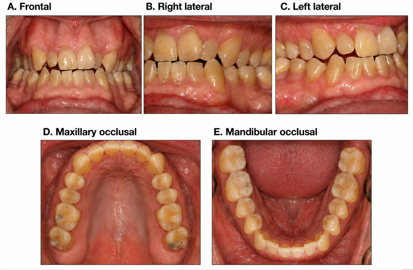
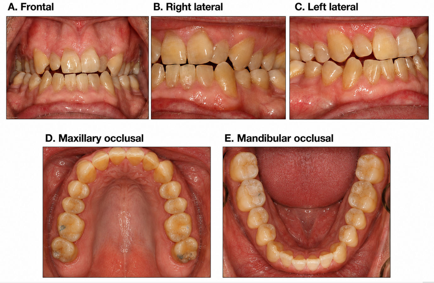
Clinical intraoral photographs documented the baseline occlusal environment, including anterior irregularity, limited anterior coupling, and posterior contact distribution. Blue articulating-paper markings recorded with 100 µm articulating paper documented static contact locations.
The articulating-paper findings were used only as a visual record of contact location. Occlusal force magnitude, timing, and right/left distribution were interpreted from T-Scan data rather than from articulating-paper mark size or intensity. (Figure 1)
T-Scan Findings
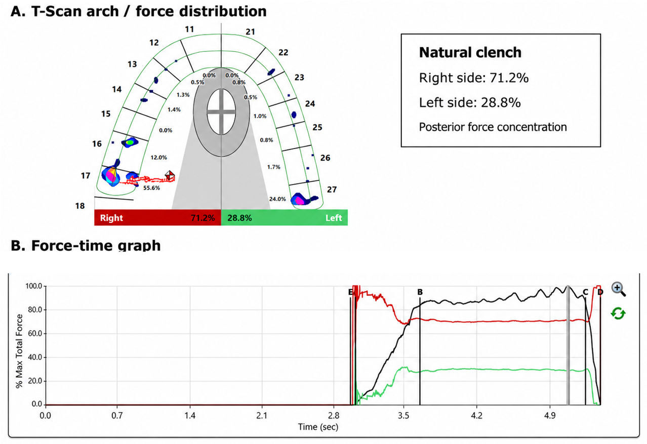
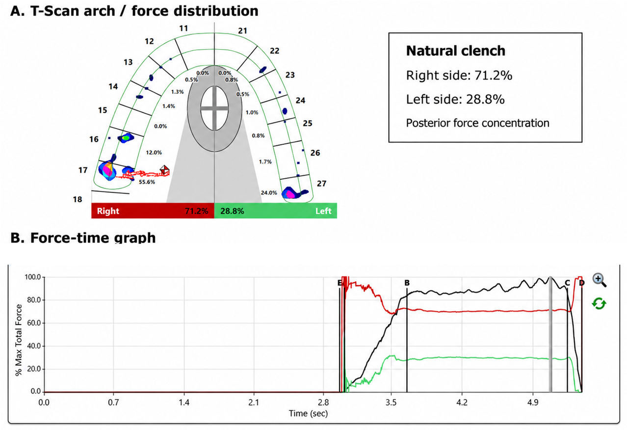
T-Scan analysis during natural intercuspation demonstrated marked right-sided force dominance and posterior force concentration. The force-time graph demonstrated early and sustained right-side dominance during the recorded clench. The occlusal load was not evenly distributed bilaterally. (Figure 2)
sEMG Findings Without and With Innobyte
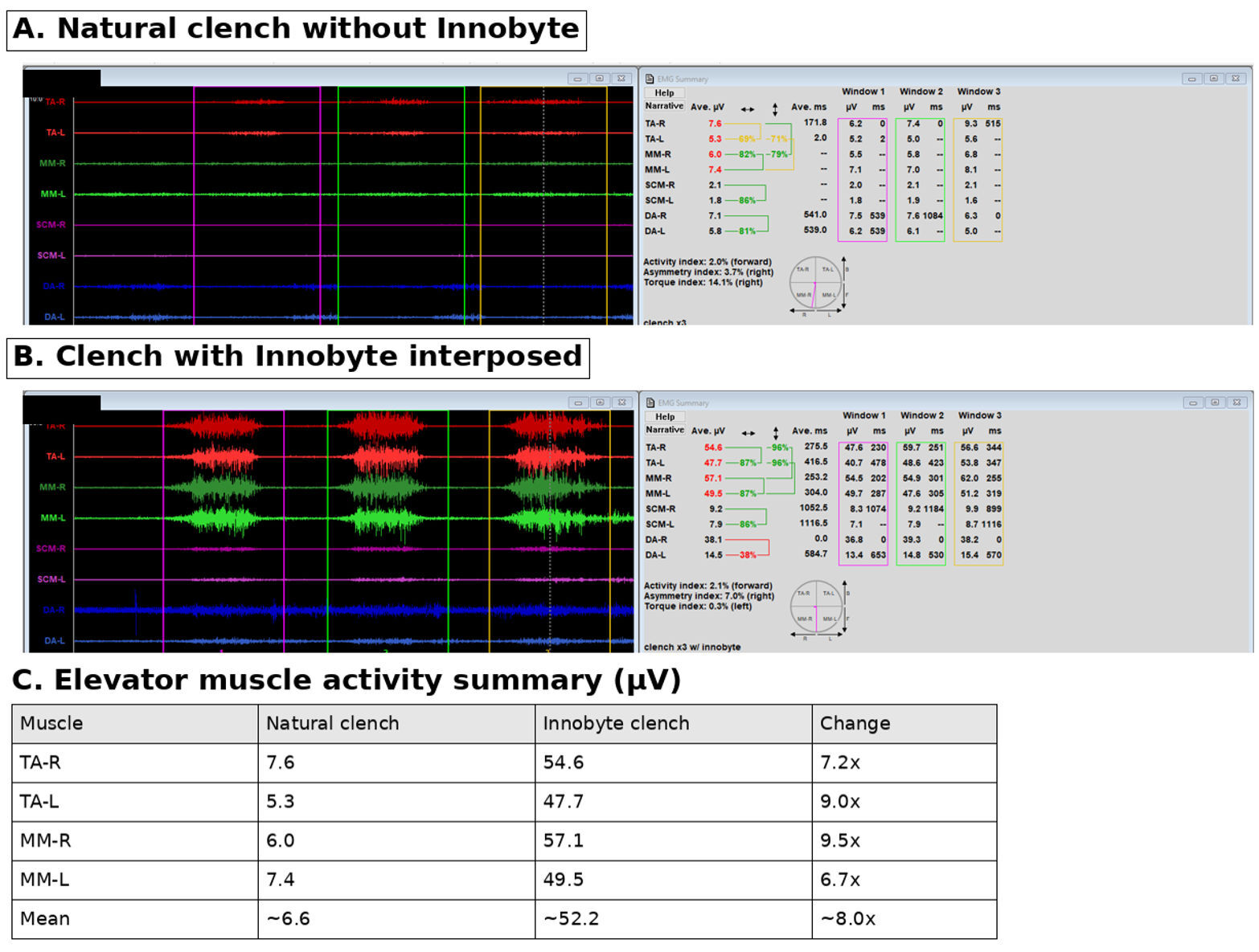
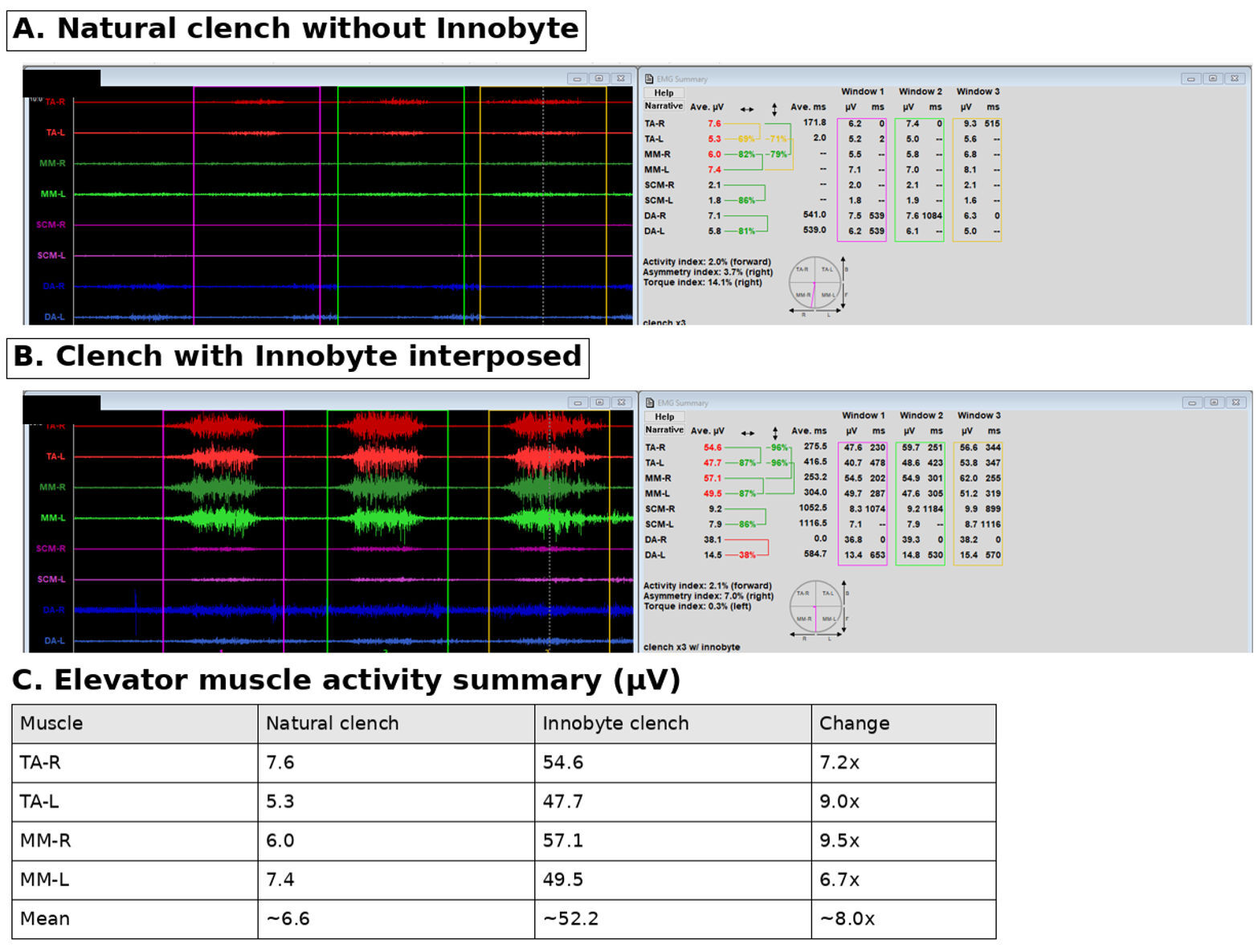
During direct clench into the natural dentition, temporalis and masseter recruitment was very low. During clench with the Innobyte interposed, elevator-muscle activity increased markedly.
Mean temporalis/masseter activity increased from approximately 6.6 µV during natural clench to approximately 52.2 µV during Innobyte-supported clench, representing an approximately eightfold increase in elevator-muscle recruitment under the altered interocclusal loading condition. (Figure 3)
Innobyte Bite-Force Findings
Innobyte testing demonstrated preserved force-generating capacity and relatively balanced right/left force distribution. Static bite force was recorded at 667 N, with 328 N on the right and 339 N on the left. The three-clench recording demonstrated 770 N total force, with 391 N on the right and 379 N on the left. These data indicate that the patient was capable of generating substantial bite force when the interocclusal loading condition was altered. (Table 2.)
Jaw Tracker and JVA Findings
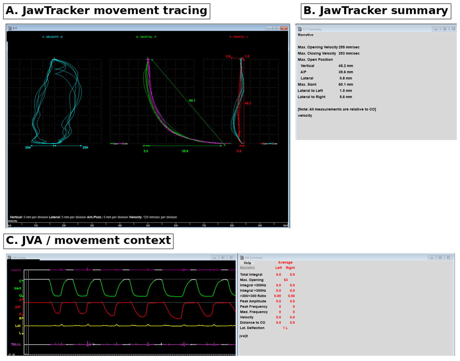
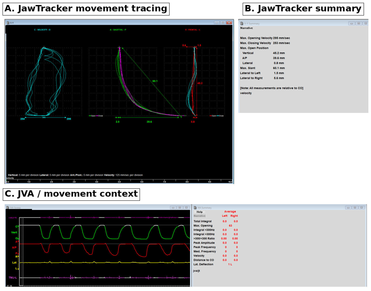
Jaw Tracker recordings demonstrated preserved mandibular opening capacity, with recorded vertical opening of approximately 45-46 mm and maximum slant values of approximately 60-63 mm. Lateral movement was mildly asymmetric across captures. Joint vibration analysis did not demonstrate a prominent vibration signature in the selected opening/closing cycle, with total integral values recorded as 0.0 bilaterally in the selected capture. This finding was interpreted cautiously and did not exclude all intra-articular pathology. (Figure 4)
Discussion
This example case illustrates the distinction between clinical diagnosis and functional characterization. The diagnosis of myofascial pain was made clinically, based on symptom history, function-related pain, and reproduction of pain during examination of the masticatory and associated cervical musculature. This is consistent with DC/TMD principles, which emphasize clinical history and standardized examination rather than electrodiagnostic testing as the basis for diagnosing myalgia or myofascial pain.1,2
The biometric findings provided additional functional information. During direct clench into the natural dentition, the patient demonstrated very low temporalis and masseter recruitment. When the Innobyte was placed between the teeth, temporalis and masseter activity increased markedly and became more organized. This indicates that the patient retained substantial elevator-muscle capacity, but that natural intercuspal loading was associated with reduced recruitment, guarding, protective inhibition, or inefficient neuromuscular output.
This finding is consistent with the broader sEMG literature showing that pain-related TMD patients may display altered masticatory muscle activity in a task-dependent manner. The literature does not support sEMG as a standalone diagnostic test for myofascial pain, but it does support its use as an adjunctive method for evaluating masticatory muscle behavior.9
The T-Scan finding provides an important mechanical correlate. Natural clench demonstrated right-heavy posterior force concentration, with 71.2% of force on the right and 28.8% on the left. (Figure 2)
This suggests that the patient’s natural intercuspal position did not provide a balanced loading platform. When the occlusal condition was changed with the Innobyte, elevator-muscle recruitment increased substantially and bite force was expressed in a more balanced right/left pattern.
The significance of this observation is not that occlusion independently diagnosed myofascial pain. Rather, the significance is that changing the occlusal environment changed function. In this patient, occlusion appeared to be a functional variable associated with masticatory muscle recruitment and force expression.
This interpretation is consistent with the cautious position required by the occlusion/TMD literature. Systematic and narrative reviews have warned against reducing TMD to a simple occlusal disease model.4,5 Experimental occlusal-interference studies also show that occlusal changes may alter motor behavior without reliably producing TMD symptoms in all subjects.6,7 These studies support a balanced interpretation: occlusal change may influence motor behavior, but occlusion should not be presented as a universal or isolated cause of myofascial pain.
A separate T-Scan/sEMG literature stream supports the clinical utility of quantifying occlusal force, timing, and associated masticatory muscle activity in selected patients. Kerstein’s early work compared disclusion time between chronic myofascial pain dysfunction syndrome patients and non-patients, helping establish a digital occlusion framework for evaluating occluso-muscular dysfunction.11 Kerstein later reported treatment of myofascial pain dysfunction syndrome by reducing lengthy disclusion time, and Kerstein and Radke subsequently reported that Disclusion Time Reduction was associated with changes in maximal clench muscle activity levels.12,13
Subsequent clinical studies further developed this approach. Thumati and colleagues reported symptom improvement after ICAGD/DTR therapy monitored with digital occlusal analysis, including a 2014 study and a later single-group interventional study with 3-year follow-up.14,15 A multicenter randomized controlled trial later used intraoral force/timing sensors synchronized with muscle physiology sensors across five dental colleges, supporting the concept that occlusal force/timing data and EMG can be evaluated together in selected occluso-muscular TMD patients.16 A later retrospective five-year survey further reported outcome data after DTR therapy in TMD patients.17
More recent ADTT literature has extended this technology-focused framework. Kunte, Sutter, and Sagwekar evaluated T-Scan 10 and BioEMG III-guided DTR therapy in patients with chronic myofascial pain symptoms, specifically examining both symptom changes and the contractile function of the anterior temporalis and masseter muscles.18 Girouard and Sutter used Innobyte to measure maximum bite force in orofacial pain patients with TMJ internal derangements under habitual bite, unadjusted orthotic, and T-Scan-adjusted orthotic conditions.24 Sutter and Girouard later reported absolute maximum bite-force changes in orofacial pain patients immediately following DTR using Innobyte.25
For the present example case, this literature supports a narrow but clinically important claim: T-Scan, sEMG, and Innobyte can be used together to characterize how occlusal loading, muscle recruitment, and bite-force expression change under specific functional conditions. These technologies do not replace DC/TMD clinical diagnostic criteria; rather, they document the functional expression of the diagnosed condition.
The example also aligns with recent calls for greater ecological validity in TMDs research. Nickel et al. emphasized that TMDs research, particularly in chronic myofascial pain, should capture objectively measured data that meaningfully reflect real functional states rather than relying only on simplified or averaged laboratory constructs.26 The present report does not claim ecological completeness, but it does document how this patient’s jaw-loading behavior changed under clinically relevant occlusal task conditions.
Innobyte adds a force-capacity dimension. Published reliability data support Innobyte as a repeatable method for measuring maximum bite force.19 In the present case example, Innobyte demonstrated that the patient was capable of generating high bite force when the interocclusal loading condition was changed. This helped distinguish low natural clench sEMG output from true muscular incapacity. The low natural-clench output was more consistent with task-dependent inhibition, guarding, altered proprioceptive input, or inefficient recruitment.
The mechanistic rationale is compatible with trigeminal sensorimotor control. Orofacial proprioception involves periodontal ligament mechanoreceptors, muscle spindles, TMJ afferents, the mesencephalic trigeminal nucleus, and trigeminal motor pathways. Lazarov described the mesencephalic trigeminal nucleus as a key structure in processing proprioceptive information from the face and oral cavity.27 Pain-adaptation models also support the concept that pain can alter motor behavior in a protective direction. Lund et al. proposed that chronic musculoskeletal pain alters motor activity through adaptive neuromuscular mechanisms rather than simple tonic hyperactivity.28 Peck et al. later described an integrated pain-adaptation model in which pain can alter recruitment strategies in ways intended to protect the sensorimotor system.29
In this case example, the Innobyte likely changed several afferent and biomechanical variables simultaneously: periodontal input, vertical dimension, occlusal contact distribution, mandibular stabilization, and load tolerance. The marked sEMG increase with Innobyte interposition is therefore consistent with a change in trigeminal sensorimotor output under a modified loading condition. Because these variables changed together, the observed response should not be attributed to a single isolated mechanism.
Jaw Tracker and JVA findings provided additional context. Mandibular opening capacity was preserved, and JVA did not show a prominent vibration signature in the recorded cycle. These findings supported the interpretation that the dominant measurable issue was not a fixed mechanical lock or vibration-dominant intra-articular disorder, but rather a myogenous and occlusal-loading functional pattern.
Taken together, the findings support the interpretation that this patient’s myofascial pain was associated with measurable occlusion-dependent neuromuscular dysfunction. The natural occlusal environment appeared to be associated with asymmetric loading and reduced elevator recruitment, while the Innobyte-supported condition allowed substantially greater and more balanced functional output.
Clinical Significance
This case example suggests that in selected patients with clinically diagnosed myofascial pain, a controlled interocclusal loading challenge may help determine whether low clench output reflects poor muscular capacity or occlusion-dependent inhibition/guarding.
The combination of sEMG, T-Scan, and Innobyte may provide useful adjunctive information for baseline functional documentation, identifying occlusion-dependent changes in muscle recruitment, distinguishing low natural-clench output from true muscular incapacity, documenting force distribution and bite-force capacity, and monitoring response to conservative therapy.
The clinical value lies in the functional observation that changing the occlusal environment changed muscle recruitment. This supports occlusion as a patient-specific functional modifier, not as a standalone diagnostic category or universal etiologic explanation.
Limitations
This single-patient review cannot establish causality or generalizable treatment efficacy.
The Innobyte changes vertical dimension, tooth contact input, periodontal afferent stimulation, mandibular posture, and load distribution. It should therefore be interpreted as a functional interocclusal challenge rather than as an isolated bite-force test. The observed sEMG change cannot be attributed to one variable alone.
sEMG is sensitive to electrode placement, skin impedance, patient effort, crosstalk, fatigue, and task standardization. T-Scan data require appropriate sensor handling and clinical interpretation. JVA findings depend on capture quality and do not exclude all possible intra-articular pathology.
The present example did not include blind assessment, repeated measures across multiple visits, asymptomatic control comparison, randomized task order, or long-term follow-up after treatment. Standardized pain scores and symptom-response follow-up were not available for this report. Future reports would be strengthened by standardized pain scales, repeated sEMG/T-Scan/Innobyte recordings, follow-up after conservative therapy, and comparison with asymptomatic or pain-free controls. Because only a single chairside session was analyzed, the findings should be viewed as hypothesis-generating and clinically descriptive rather than as evidence of generalized treatment efficacy.
Conclusion
This example was designated as myofascial pain, a non-specific TMD, clinically using history and examination findings consistent with DC/TMD principles but without any indication of an etiology. sEMG, T-Scan, Jaw Tracker, JVA, and Innobyte did not replace the clinical diagnosis. Instead, they demonstrated that masticatory muscle recruitment and force expression changed substantially when the occlusal loading condition was modified.
Natural clench was associated with low elevator-muscle recruitment and asymmetric posterior occlusal force concentration. Innobyte-supported clench produced markedly increased temporalis/masseter activity and balanced high-force output. Jaw Tracker demonstrated preserved mandibular opening capacity, and JVA did not demonstrate a prominent vibration signature in the selected cycle.
These findings support the interpretation that, in this patient, occlusal loading appeared to act as a functional modifier of neuromuscular behavior and may have contributed to protective inhibition, guarding, inefficient recruitment, and persistence or expression of myofascial pain.
Patient Consent
Was obtained when the patient was tested, and was applicable for this de-identified retrospective clinical review. The manuscript was prepared from routine clinical records and contains no information intended to identify the patient. All figures and biometric screenshots should remain fully de-identified before submission.
Ethics Statement
This manuscript describes a single retrospective clinical case example that is based on routine diagnostic assessment and de-identified clinical data. No experimental intervention was performed for any purpose. Formal institutional review board approval was not sought for this de-identified case example.
Conflict of Interest
The author declares no conflict of interest.
Funding
No external funding was received for preparation of this manuscript.
Author Contributions
PG performed the clinical evaluation, interpreted the biometric recordings, conceptualized the review, prepared the manuscript, and approved the final version. This statement should be revised if additional authors are added.
Data Availability
De-identified supporting clinical data may be available from the corresponding author with all reasonable requests, subject to applicable privacy and professional record requirements.