


_in_figure_11__.tiff)
The International Headache Society characterizes Headache Disorders as primary or secondary.1 Primary headaches are classified as Tension Headache, Migraine, Cluster, and Exertional, while secondary headaches occur in response to a disease pathology resultant from exogenous factors (i.e., a vascular disorder, head injury, brain tumor, or hemorrhage).
Migraine is defined by the International Classification of Headache Disorders (ICHD) as headache attacks lasting for 4 - 72 hours, that are accompanied by nausea, photophobia and phonophobia, or both. Migraine headache is classically unilateral, pulsating, and of moderate to severe intensity, which is aggravated by physical activity.2
A Migraine attack is thought to be a complex, multifactorial disorder of the brain. It is classically divided into four clinical phases:
-
Premonitory symptoms
-
Aura
-
Headache
-
Postdrome phase.
These phases can occur in a linear sequential order or significantly overlap with the latter being more common. However, the actual migraine etiology remains in question.
The Premonitory Symptoms include concentration impairment, yawning, mood change, and homeostatic changes in eating behavior and fluid balance that are typically seen within 72 hours of headache onset but can been seen any time before the onset of headache. The trigger factors described by patients with migraines can be sleep deprivation, hunger and/or fasting, bright lights or loud sounds may initiate premonitory symptoms of an ongoing attack.
The Aura Phase can include transient, but fully reversible neurological deficits, often localized to one side of the head. Aural symptoms develop over 5 minutes, and last between 5- 60 minutes. Migraine aura is experienced by about one-third of migraine patients, and can act as predictor of the imminent headache, commonly preceding the headache phase, but can well last into the headache phase itself.
Visual aura is the most common aura phase symptom, observed in 90% of the cases. It can be either positive with fortification spectra – zig zag lines or flashing lights across visual fields, or negative with scotoma, or both. Aura motor deficits like those seen with familial hemiplegic migraines include sensory or speech deficits that often last longer than does visual aura.
The Headache Phase is clinically diagnosed using the simplified diagnostic headache criteria.1
The Postdrome Phase is when the headache phase is followed by tiredness, decreased concentration, and neck stiffness.
The cause of Migraine has been postulated to be vascular by Wolff in 1948,2 due to vasodilation and excessive pulsation of the branches of external carotid artery that explained the pulsatile and throbbing nature of the headache. Studies by Olsen et al, and Cutter using a xenon inhalation method of brain imaging SPECT; (Single Photon Emission Tomography), PET scan, and perfusion weighted MRI during an attack, seemed to support the idea of a vascular etiology, but have not yet proven it.3
Observations made by Leao using rats led to what became known as Cortical Spreading Depression (CSD), that also attempted to explain the origin of headache in migraine.3 A noxious stimulus applied to rat cortex induced a vasoconstriction that slowly inhibited the electrical activity of many cortical neurons. However, it is not common for a migraine patient to have a history of receiving a noxious injection into their cortex.
An alternative theory by Moskowitz3 hypothesized that neural mechanisms in the Trigemino-vascular complex (TVC) were the basis of aura and headache in migraine patients. Both intra and extra cranial vasculature innervated by unmyelinated trigeminal nerves (mostly from the ophthalmic branch (V1), and to a lesser extent by the maxillary (V2) and mandibular (V3) branches) when activated, released substance P, Calcitonin gene related peptide (CGRP), and other peptides into blood vessel walls, which sensitized the trigeminal system to cranial vessel pulsations to promote an inflammatory response.
Dental Occlusion as a cause of migraine has been minimally noted in medical literature. However, when PubMed was searched for "Headache AND “dental splint OR dental orthotic OR dental appliance”, 213 listings were found. In one such Case Report a patient diagnosed with migraine and aura and Temporomandibular Joint and masseter muscle pain, was treated with a dental appliance that eliminated the headache, the visual aura, and reduced the other symptoms.4 This Case Report supports the findings of some allergists who have reported a correlation exists between TMD, occlusion and headache.
However, multiple studies involving muscular TMD patients have reported computer-guided occlusal adjustments can lessen chronic headaches.5–18 Chronic orofacial pain in the Temporomandibular joint region is often accompanied by frequent temporal headaches, in combination with neck pain, ear pain, chewing fatigue, clenching and grinding habits, morning muscle stiffness, limited mouth opening, with clicking or popping sounds in Temporomandibular joints.19 Chronic orofacial pain occlusal adjustment studies have shown improvement of headache symptoms, whereby the treated groups showed a strong relation between occlusal adjustments and headache improvements.7,20–22
The T-Scan computerized occlusal analysis system (Tekscan, Inc. S. Boston, MA, USA) digitally analyses the occlusion quantitatively, by reporting real-time occlusal contact sequencing measurements that play as a video in dynamic force movies. Reducing the Disclusion Time of all molars and premolars to < 0.5 seconds per excursion has been shown to reduce muscle hyperactivity levels and its related symptoms, including temporal headaches pre to post treatment.5,7,11,12,22 The Disclusion Time is reduced using a T-Scan guided and measured occlusal adjustment procedure known as Immediate Complete Anterior Guidance Development (ICAGD),11,16 wherein excursive occlusal interferences are removed selectively until the Disclusion Time value becomes < 0.5 seconds per excursion. This lessened masticatory muscle activity has positively improved headache in patients treated occlusally.13,17–19,22
To date there are no published headache-specific occlusal adjustment studies. As such, the Specific Aims of this study were to perform a Disclusion Time Reduction study with headache and migraine patients as a required population criterion, to observe any possible treatment effects that Disclusion Time Reduction offers chronic headache patients.
Method and Material
120 patients from Rajarajeshwari Dental College and Hospital, at Rajiv Gandhi University of Health Sciences, filled out a Headache questionnaire that qualified them for possible study participation. The questions inquired about their history of headache symptoms, treatments they received in the past, and the frequency and intensity of their common daily symptoms. Each potential subject also completed a Beck Depression Inventory-II questionnaire (BDI –II),23–25 which scored their level of emotional depression related to living with chronic headaches.
The Inclusion criteria were
-
A history of reported chronic headaches that were unsuccessfully lessened by prior treatments.
-
A fully dentulous state of at least 28 teeth
-
Near normal occlusal relations with molars and premolars in contact in right and left excursions.
-
Angles Class I and Class III subjects with guiding teeth that were either in contact, or near to contact.
Note: Not every headache patient is a candidate for DTR; only those specified within these Inclusion criteria, and those not excluded by other criteria.
The Exclusion criteria were
-
Severe Class II and Class III malocclusions and large anterior open occlusions, where anterior guidance contacts did not exist
-
A previous history of head trauma
-
Patients who had undergone prior occlusal adjustment treatment
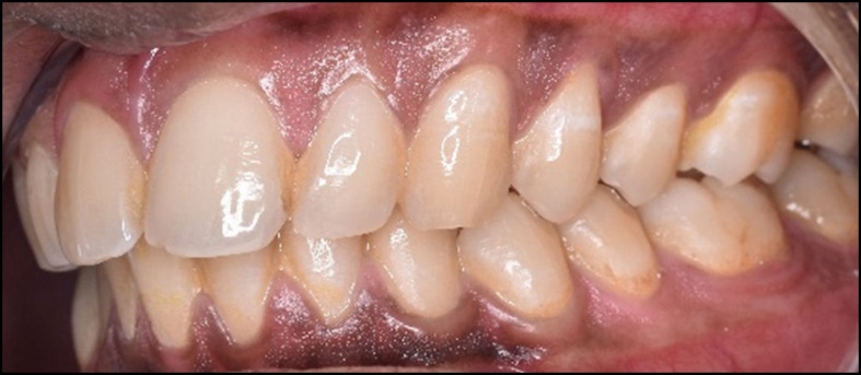
Ultimately, the 120 selected cases all demonstrated chronic headaches, medium to high Beck Depression scores, presented with Angles Class 1 skeletal relations and 28 teeth, with many subjects experiencing both muscular and Temporomandibular Joint symptoms in addition to headaches. Fifty-six of the 120 patients had undergone prior Orthodontic treatment. A representative headache subject is depicted in Figures 1-3.


All subjects filled out an informed consent that stated the participant subjects would receive occlusal adjustment treatment. The protocol was reviewed and approved by the IRB of the Health University of Rajiv Gandhi University of Health Sciences (RGUHS).
TREATMENT PROCEDURE
The T-Scan 10 synchronized to the BioEMG III (BioResearch Associates, Inc. Milwaukee, WI USA) was used to record Maximum Intercuspation (MIP), and the right and left excursions of all subjects. Subjects closed into their MIP firmly, and either opened and re-closed into MIP again (Figure 4), or held their teeth together for 1-3 seconds, and then commenced a right or left excursion, until only anterior teeth were in contact. This specific recording method assures that high quality Disclusion Time and EMG data are obtained from all subjects.26

Each subject’s pre-treatment Disclusion Time values per excursion and the excursive electromyography levels were compared to the same subject’s post ICAGD coronoplasty excursive Disclusion Time values and EMG levels. See Figures 4 to 6


All of the Day 1 pre-treatment Disclusion Time/EMG data was compared to Day 1 post-treatment Disclusion Time/EMG data, and to all subsequent dates of treatment or recall (1 week, 1 month, 3 months 6-months).
ICAGD was then commenced with excursive contact adjustments. The subjects’ teeth were air dried. The patients were then asked to close into their Maximum Intercuspal Position (MIP) with articulating paper (Bausch, Arti-Fol® Red, 8μ, Germany) interposed, and then to commence a right mandibular excursion out to edges of their canine teeth, then slide back into MIP, and then make a left mandibular excursion out to the edges of the left side canine teeth, and back into MIP.
The pre-treatment T-Scan 10/BioEMG III recordings guided the areas to correct, where prolonged excursive frictional contacts were detected in the resultant articulating paper markings (inclined plane linear contact patterns) (Figure 7). These excursive contacts were then eliminated on all involved surfaces using pear shaped finishing burs (Mani Dia-Burs, Japan ISO no-237/021) bilaterally, leaving the central fossa, cusp tip, and the marginal ridge contact points intact.

ICAGD was complete when:
-
All Glickman Class I, II, and III lateral excursive interferences27 had been visually removed
-
Disclusion of all posterior teeth in the right and left excursions was visible with the patient experiencing easier lateral movements than pre-ICAGD
-
All lace-like linear contacts had been removed
-
The new MIP closure contacts mostly located on cusp tips, in fossae, or on marginal ridges (Figure 8)
-
New recordings of each excursion verified that the Disclusion Times had been reduced to < 0.5 seconds per excursion (Figures 9 and 10).


At day 7 post ICAGD, each subject filled out new symptom questionnaires, and made new habitual closure into MIP and excursive T-Scan 10/BioEMG III recordings. This new data guided new MIP closure and excursive refinement adjustments, that were followed by additional recordings to document the improved closure and excursive EMG levels present.
At 1, 3, and 6 months, repeat measurements were obtained and new symptom questionnaires were answered, but no further occlusal treatment was rendered.
METHOD OF SYMPTOM ASSESSMENT
Patient self-assessment with repeated BDI-II indices and verbally scored headache frequency and intensity questionnaires were used pre-ICAGD, and at all dates of treatment or biometric measurement. Each questionnaire acted as its own assessment for each date that followed treatment Day 1.
The Disclusion Time values pre and post ICAGD, and the excursive EMG levels pre and post ICAGD were subjected to the Student’s paired t-Test (Alpha = 0.05). The BDI-II Scores, Headache Pain Scores, Headache Frequency Analysis, Functional Restrictions, and the Frequency of Painful Symptoms were all subjected to the non-parametric Wilcoxon Signed-Rank Test for paired data (Alpha = 0.05).
RESULTS
The Disclusion Times were significantly reduced (p < 0.05) at each time point, except between the week 1 to 1 month period for the left excursions (p = 0.0906). See Table 1. The DT reductions were most dramatic between pre-treatment and week 1 but continued to reduce up to 6 months post treatment.
The Beck Depression Inventory – II scores significantly reduced dramatically between the pre-treatment time point and 1 week post treatment (p = 0.00001). Although there was no significant change at 1 month, at 3 and 6 months significant BDI-II score reductions were observed. See Table 2.
The Pain Scale data revealed very large significant reductions 1 week post treatment. Additional, less dramatic but still significant reductions occurred at 1 month and 3 months. A slight further reduction at 6 months did not reach statistical significance. See Table 3.
Table 4 tracked the frequency of the symptoms listed in Table 3. Very dramatic and significant frequency reductions of all symptoms occurred one week after treatment up to 1 month. Non-significant reductions in symptom frequency were seen at 3 and 6 months post-ICAGD.
The Functional Restrictions, see Table 5, underwent significant reductions between pre-treatment and 1 week post ICAGD (p = 0.0027), with further significant reductions occurring between 1 week and 3 months post ICAGD (p = 0.04026).
Table 6 focused solely on the frequency of painful symptoms, where a large and significant painful symptom frequency reduction of the painful symptoms occurred from pre-treatment to 1 week post ICAGD (p < 0.0027). At each time point thereafter, further significant frequency reductions in painful symptoms were observed.
Table 7 shows the significant differences in EMG means per muscle, between the C and D time moments during the excursive movements from before ICAGD to 6 months after the Disclusion Times were markedly shortened at Day 1 after ICAGD treatment. The combined left and right excursive EMG activity of the bilateral masseter and anterior temporalis muscles at “D” (complete posterior disclusion bilaterally), significantly and progressively reduced at each time point (p < 0.05), with the exception of the pre-treatment to 1-week time period.
The EMG levels at “C” (end of MIP before excursive commencement) increased at each timepoint because the subjects became more comfortable intercuspating forcefully, as closure interferences were no longer present after ICAGD. At the same time, the EMG levels at “D” declined, because subjects required much less muscular effort to make their lateral excursive movements, with the excursive friction removed from their occlusal surfaces by ICAGD. The adaptation was gradual, as the mean differences in EMG levels between C and D increased over the 6 months of study observation.
Discussion
The Results of this ICAGD interventional clinical study do not exactly corroborate the findings of previous occlusal adjustment headache studies, as solely headache DTR/ICAGD occlusal adjustment studies have not been previously undertaken. However, the Results of this ICAGD headache study are substantially similar to previously published muscular TMD treatment studies,5–19 which tracked headache symptom intensities and frequencies and their emotional impact on subjects that experienced frequent headache as a symptom.7,18,19,27 Prior studies found painful headaches improved within one week after computer-guided ICAGD was rendered and the excursive Disclusion Times were properly shortened, that continued to improve as further treatment and time occurred out from the Day 1 ICAGD treatment.5,7,11,12,22 This study’s statistically analyzed Results indicate the same trend occurred, in that most of the 120 headache subjects experienced marked and rapid headache symptom pain intensity and frequency reductions, 1 week after the Disclusion Times were shortened with ICAGD (Table 1), which continued throughout the remainder of the 6 months period of subject observation (Tables 2 - 6).
Of note is that the subjects’ Beck Depression Inventory scores (Table 2) dropped significantly after ICAGD was rendered, as the subjects’ headache frequency and pain levels lessened. The group’s emotional improvements were concurrent with the lessening of their chronic headache conditions. This finding corroborates prior Disclusion Time Reduction studies that found when chronic muscular TMD pain was reduced in frequency and intensity, ongoing emotional depression was quickly reversed into emotional well-being, restoring improved emotional quality of life.7,18,19,28
Table 3 clearly indicates that ICAGD occlusal treatment markedly lessened the frequency of all types of headaches, including Migraine, with statistical significance. Migraine-type headache caused by the occlusion is not well understood in the medical profession, as the motor neurology involved from the pulp and PDL fibers of posterior teeth has not been directly tied to the etiology of migraine. Moskowitz3 hypothesized that all of the neural mechanisms in the Trigeminovascular complex (TVC) were the basis of aura and headache in migraine patients. This theory details that the meningeal nociceptor neurons that innervate the cerebral blood vessels, whose cell bodies are located in the trigeminal ganglion, are consequentially activated within the brainstem, which leads to a repetitive blood vessel pulsation type headache. However, the results of this study suggest that the same occluso-neurologic physiology (absent of a vascular component) that has been well-described in a number of prior muscular TMD Disclusion Time Reduction treatment studies,5–19,22,29 is at play in the true etiology of many chronic headache conditions.
The molar and premolar pulp and Periodontal Ligament (PDL) mechanoreceptors have unique neuroanatomy, in that despite being Peripheral Nerves, they synapse initially inside the Central Nervous System, entering the CNS directly via the Mesencephalic Nucleus, and without synapsing there, travel on further to the Trigeminal Motor Nucleus bilaterally. These afferent fibers also travel directly to the Reticular Formation,30–32 which is an important primal brainstem center that controls functions like sleep, gastrointestinal activity, arousal, respiration rate, heart rate and an important regulator of the autonomic system. See Figure 11.
_of_the_molar_and_premolar_pulp_and_pdl_mechanorecept.jpeg)
Within the Trigeminal Motor Nucleus, the pulpal and PDL mechanoreceptor fibers make their first synapse with the efferent trigeminal motor fibers to the muscles of mastication that include the temporalis (the headache muscle), the masseter, the internal and lateral pterygoids, the tensor tympani, the tensor veli palatini, the mylohyoid, and the anterior belly of the digastric muscles, all of which comprise the muscles of swallowing.30 See Figure 12.
_in_figure_11__.tiff)
A Probable Explanation for how malocclusion can produce headache
Tooth contact that compresses and flexes teeth under loading, then creates the conduit through which much electrical activity from posterior teeth is pushed back into and out of the brain, in an ongoing way, day after day after day, with each chew, clench and swallow that a patient performs. These opposing posterior tooth compressions result in compressions of the same occluding molar and premolar pulp and PDL mechanoreceptor fibers, that via their 1st synapse within the Trigeminal Motor Nucleus, contracts the four muscles of mastication (including the temporalis), and the other individual palate, ear, and floor of the mouth muscles, thousands of times per day. These ongoing contractions can make the Swallowing muscles chronically hyperactive, which leads to a host of symptoms and pain conditions seen in TMD patients. With respect to “headache” specifically, this occlusion-activated neural flexure and compressive mechanism hyper-functions the temporalis muscles to become ischemic, painful and dysfunctional, which then appears clinically as chronic and repetitive headache pain.
By removing the prolonged occlusal surface friction with ICAGD to shorten the Disclusion Time (DT), the neural mechanism is quieted by drastically reducing the electrical pushback into and out of the CNS, that allows for the ischemia to be metabolically resolved and to not recur, despite the fact that patients chew, eat, and swallow thousands of times per day. The absence of the frictional occlusal contacts that caused the neural response controls the neural mechanism, despite the neuroanatomy still being in place. A shortened DT has been shown repeatedly to resolve chronic muscular pain and dysfunction in TMD patients.5–19,22,29
In this headache-specific study, the same shortened Disclusion Time was able to resolve many differing types of chronic headaches rapidly and effectively. This was attributed to the statistically significant differences in the excursive muscle activity levels between C and D (Table 7). The masseter and anterior temporalis excursive movement muscle activity significantly and progressively reduced (between C& D; compare Figures 5 and 6 with Figures 9 and 10), illustrating the physiologic effect that short Disclusion Time created from within the subject pool’s neuroanatomy.
While lateral excursive movements are certainly not functional movements, it has been shown previously that removing lateral excursive interfering posterior contacts can improve masticatory function.6 This is evidence that the interference to lateral excursive movements may also be the occlusal interferences that can affect mastication.
Limitations
The absence of a control headache subject group is a drawback of any treatment study, as some Placebo effect cannot be entirely ruled out. However, aside from pharmaceutical (medication) research, true placebo control groups are difficult to manage ethically and physically, when an irreversible dental treatment is involved. Since one of the Inclusion criteria was “unsuccessful previous treatment”, the likelihood of a spontaneous or exaggerated placebo effect affecting this study’s Results seems low. And, the repeated use of verbal rating scales for pain intensity, symptom frequency and functional restriction questionnaires tracked over time, as to how the 120 subjects responded to the ICAGD treatment aimed at their pre-treatment headache condition, allowed the 120 subjects to act as their own controls. They were recalled for multiple dates of observation and were given many opportunities to respond to multiple questionnaires about their life’s conditions from before to after ICAGD treatment. The statistically consistent and marked symptom frequency and intensity declines following ICAGD seen in Tables 2-6, suggest that there was an efficacious treatment effect shared by most subjects.
Conclusions
Migraine has been considered a complex, potentially heritable, variable disorder of brain function that may have a significant dental occlusion component. Most of the 120 headache subjects experienced marked and rapid headache symptom frequency and pain level reductions, following undergoing the ICAGD treatment, which included Migraine as a responding headache subset. The Disclusion Time changes lessened the ongoing temporalis muscle contractions, that in turn relieved the chronically hyperactive muscular state, while also alleviating the ischemia, which ultimately reduced the frequency and intensity of the subjects’ headaches. The findings of this study indicate the medical community that treats chronic headache conditions should consider dental occlusion and long Disclusion Time as potential differential diagnoses in their chronic temporal headache and migraine patients.
Funding statement
No funding was received.
Potential Conflict Statement
Dr Kerstein is a consultant to Tekscan, Inc., but receives no monitory benefit from sales. Mr. Radke is Chairman of the Board of BioResearch Associates, Inc.